{"title":"汞和硒在水俣病患者脑、小脑、肝和肾中的定位。","authors":"Masumi Marumoto, Mineshi Sakamoto, Kohji Marumoto, Shozo Tsuruta, Yoshihiro Komohara","doi":"10.1267/ahc.20-00009","DOIUrl":null,"url":null,"abstract":"<p><p>Minamata disease is a methylmercury poisoning caused by consumption of marine food contaminated by man-made methylmercury environmental pollution, and its most prominent feature is marked pathological changes in the central nervous system. Morphological alterations are less pronounced in the liver and the kidney, although their mercury levels are higher than those of the brain. In marine mammals, methylmercury is known to be easily converted to inorganic mercury and it combines with selenium forming mercury selenide, which may counteract the toxicity of mercury. However, little is known about the formation of mercury and selenium complex in human organs. In the present study, we examined the cerebrum, cerebellum, liver, and kidney of a Minamata disease case to study the mercury and selenium localization using electron probe microanalysis. Our results indicated the mercury and selenium localization in the specified tissue of the brain, liver, and kidney such as glial cells, Kupffer cells, and renal tubules.</p>","PeriodicalId":6888,"journal":{"name":"Acta Histochemica Et Cytochemica","volume":"53 6","pages":"147-155"},"PeriodicalIF":1.6000,"publicationDate":"2020-12-25","publicationTypes":"Journal Article","fieldsOfStudy":null,"isOpenAccess":false,"openAccessPdf":"https://www.ncbi.nlm.nih.gov/pmc/articles/PMC7785461/pdf/","citationCount":"9","resultStr":"{\"title\":\"Mercury and Selenium Localization in the Cerebrum, Cerebellum, Liver, and Kidney of a Minamata Disease Case.\",\"authors\":\"Masumi Marumoto, Mineshi Sakamoto, Kohji Marumoto, Shozo Tsuruta, Yoshihiro Komohara\",\"doi\":\"10.1267/ahc.20-00009\",\"DOIUrl\":null,\"url\":null,\"abstract\":\"<p><p>Minamata disease is a methylmercury poisoning caused by consumption of marine food contaminated by man-made methylmercury environmental pollution, and its most prominent feature is marked pathological changes in the central nervous system. Morphological alterations are less pronounced in the liver and the kidney, although their mercury levels are higher than those of the brain. In marine mammals, methylmercury is known to be easily converted to inorganic mercury and it combines with selenium forming mercury selenide, which may counteract the toxicity of mercury. However, little is known about the formation of mercury and selenium complex in human organs. In the present study, we examined the cerebrum, cerebellum, liver, and kidney of a Minamata disease case to study the mercury and selenium localization using electron probe microanalysis. Our results indicated the mercury and selenium localization in the specified tissue of the brain, liver, and kidney such as glial cells, Kupffer cells, and renal tubules.</p>\",\"PeriodicalId\":6888,\"journal\":{\"name\":\"Acta Histochemica Et Cytochemica\",\"volume\":\"53 6\",\"pages\":\"147-155\"},\"PeriodicalIF\":1.6000,\"publicationDate\":\"2020-12-25\",\"publicationTypes\":\"Journal Article\",\"fieldsOfStudy\":null,\"isOpenAccess\":false,\"openAccessPdf\":\"https://www.ncbi.nlm.nih.gov/pmc/articles/PMC7785461/pdf/\",\"citationCount\":\"9\",\"resultStr\":null,\"platform\":\"Semanticscholar\",\"paperid\":null,\"PeriodicalName\":\"Acta Histochemica Et Cytochemica\",\"FirstCategoryId\":\"99\",\"ListUrlMain\":\"https://doi.org/10.1267/ahc.20-00009\",\"RegionNum\":4,\"RegionCategory\":\"生物学\",\"ArticlePicture\":[],\"TitleCN\":null,\"AbstractTextCN\":null,\"PMCID\":null,\"EPubDate\":\"2020/12/19 0:00:00\",\"PubModel\":\"Epub\",\"JCR\":\"Q4\",\"JCRName\":\"CELL BIOLOGY\",\"Score\":null,\"Total\":0}","platform":"Semanticscholar","paperid":null,"PeriodicalName":"Acta Histochemica Et Cytochemica","FirstCategoryId":"99","ListUrlMain":"https://doi.org/10.1267/ahc.20-00009","RegionNum":4,"RegionCategory":"生物学","ArticlePicture":[],"TitleCN":null,"AbstractTextCN":null,"PMCID":null,"EPubDate":"2020/12/19 0:00:00","PubModel":"Epub","JCR":"Q4","JCRName":"CELL BIOLOGY","Score":null,"Total":0}
Mercury and Selenium Localization in the Cerebrum, Cerebellum, Liver, and Kidney of a Minamata Disease Case.
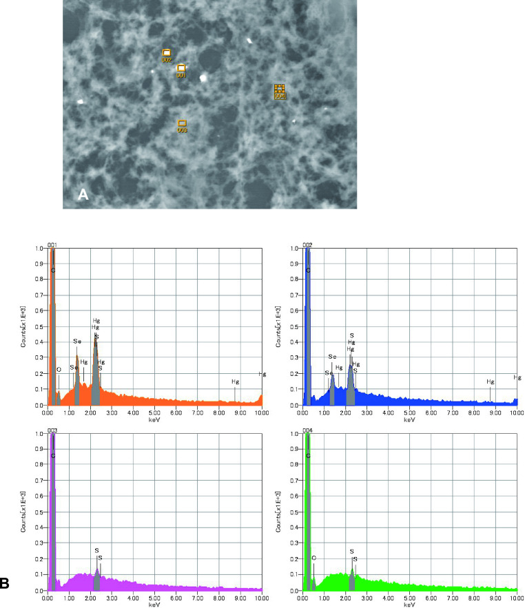
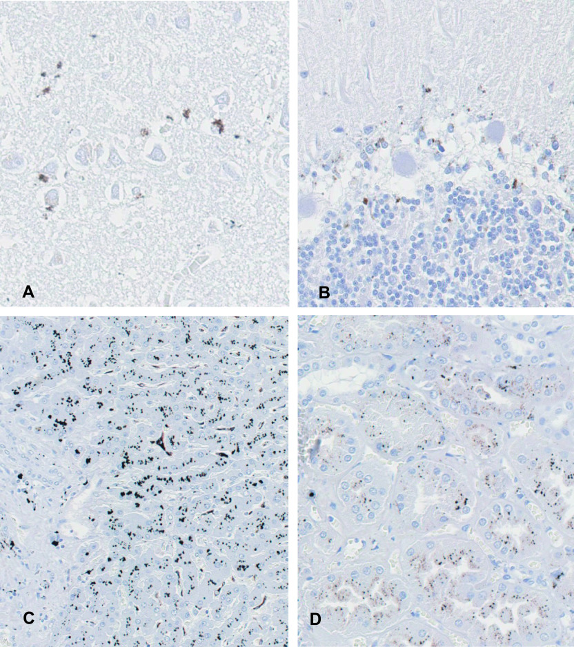
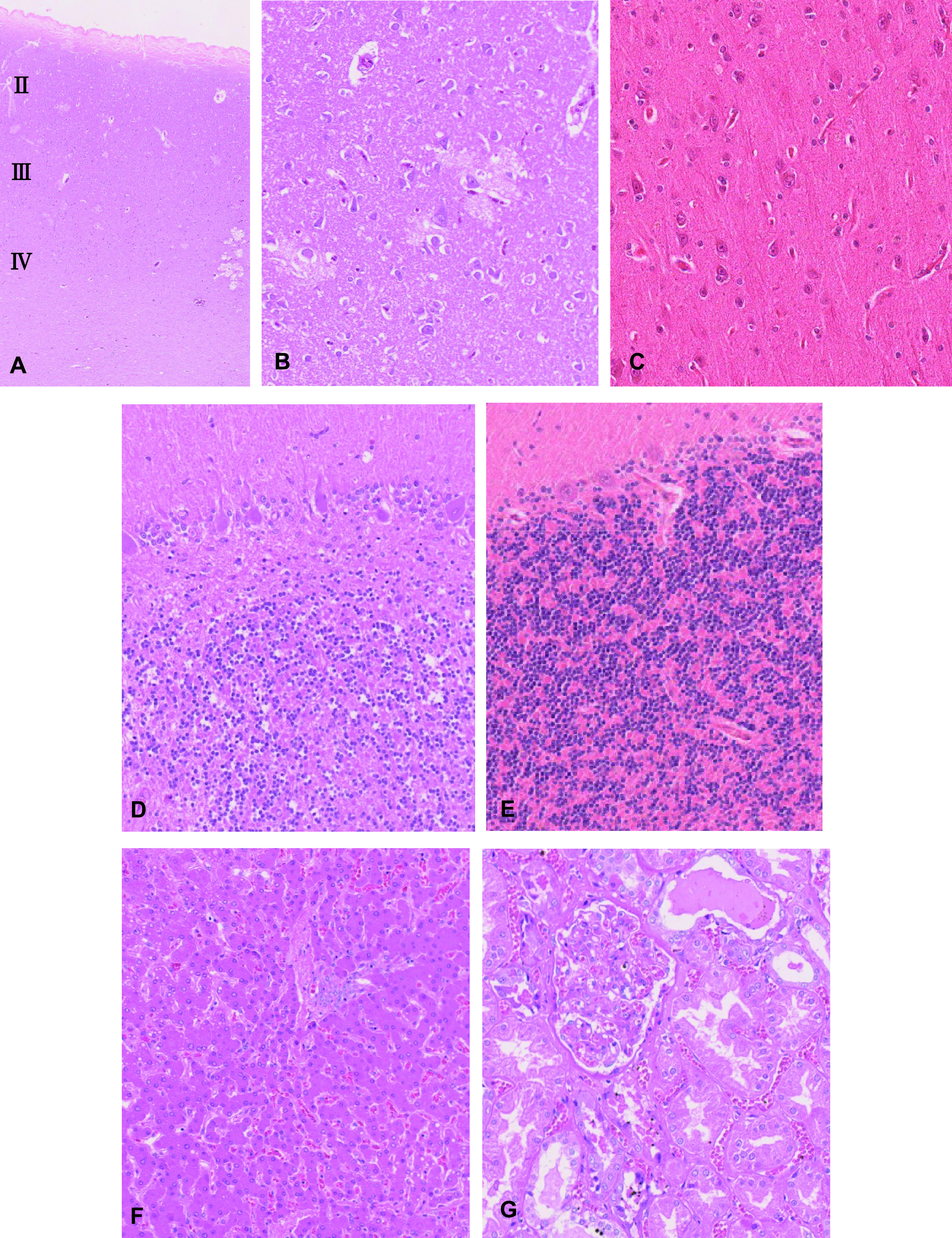
Minamata disease is a methylmercury poisoning caused by consumption of marine food contaminated by man-made methylmercury environmental pollution, and its most prominent feature is marked pathological changes in the central nervous system. Morphological alterations are less pronounced in the liver and the kidney, although their mercury levels are higher than those of the brain. In marine mammals, methylmercury is known to be easily converted to inorganic mercury and it combines with selenium forming mercury selenide, which may counteract the toxicity of mercury. However, little is known about the formation of mercury and selenium complex in human organs. In the present study, we examined the cerebrum, cerebellum, liver, and kidney of a Minamata disease case to study the mercury and selenium localization using electron probe microanalysis. Our results indicated the mercury and selenium localization in the specified tissue of the brain, liver, and kidney such as glial cells, Kupffer cells, and renal tubules.
期刊介绍:
Acta Histochemica et Cytochemica is the official online journal of the Japan Society of Histochemistry and Cytochemistry. It is intended primarily for rapid publication of concise, original articles in the fields of histochemistry and cytochemistry. Manuscripts oriented towards methodological subjects that contain significant technical advances in these fields are also welcome. Manuscripts in English are accepted from investigators in any country, whether or not they are members of the Japan Society of Histochemistry and Cytochemistry. Manuscripts should be original work that has not been previously published and is not being considered for publication elsewhere, with the exception of abstracts. Manuscripts with essentially the same content as a paper that has been published or accepted, or is under consideration for publication, will not be considered. All submitted papers will be peer-reviewed by at least two referees selected by an appropriate Associate Editor. Acceptance is based on scientific significance, originality, and clarity. When required, a revised manuscript should be submitted within 3 months, otherwise it will be considered to be a new submission. The Editor-in-Chief will make all final decisions regarding acceptance.
 求助内容:
求助内容: 应助结果提醒方式:


