{"title":"米诺环素减轻Zitter突变大鼠黑质活化小胶质细胞簇形成和年龄依赖性多巴胺能细胞死亡。","authors":"Daisuke Taguchi, Ayuka Ehara, Taro Kadowaki, Shin-Ichi Sakakibara, Kazuhiko Nakadate, Koichi Hirata, Shuichi Ueda","doi":"10.1267/ahc.20-00022","DOIUrl":null,"url":null,"abstract":"<p><p>Microglial activation is a component of neurodegenerative pathology. Here, we examine whether activated microglia participate in age-related dopaminergic (DA) cell death in the substantia nigra pars compacta (SNc) of the zitter (<i>zi/zi</i>) rat, a mutant characterized by deletion of the attractin gene. Confocal microscopy with double-immunohistochemical staining revealed activated microglia-formed cell-clusters surrounding DA neurons in the SNc from 2 weeks after birth. An immunoelectron microscopic study showed that the cytoplasm of activated microglia usually contains phagosome-like vacuoles and lamellar inclusions. Expression levels of the pro-inflammatory cytokines <i>interleukin-1β</i> (<i>IL-1β</i>), <i>tumor necrosis factor-α</i> (<i>TNF-α</i>) and <i>inducible nitric oxide synthase</i> (<i>iNOS</i>) were increased in the midbrain of 2-month-old <i>zi/zi</i> rats. Chronic treatment with the anti-inflammatory agent minocycline altered the morphology of the microglia, reduced cluster formation by the microglia, and attenuated DA cell death in the SNc, and reduced the expression of <i>IL-1β</i> in the midbrain. These results indicate that activated microglia, at least in part and especially at the initial phase, contribute to DA cell death in the SNc of the <i>zi/zi</i> rat.</p>","PeriodicalId":6888,"journal":{"name":"Acta Histochemica Et Cytochemica","volume":"53 6","pages":"139-146"},"PeriodicalIF":1.6000,"publicationDate":"2020-12-25","publicationTypes":"Journal Article","fieldsOfStudy":null,"isOpenAccess":false,"openAccessPdf":"https://www.ncbi.nlm.nih.gov/pmc/articles/PMC7785462/pdf/","citationCount":"1","resultStr":"{\"title\":\"Minocycline Alleviates Cluster Formation of Activated Microglia and Age-dependent Dopaminergic Cell Death in the Substantia Nigra of Zitter Mutant Rat.\",\"authors\":\"Daisuke Taguchi, Ayuka Ehara, Taro Kadowaki, Shin-Ichi Sakakibara, Kazuhiko Nakadate, Koichi Hirata, Shuichi Ueda\",\"doi\":\"10.1267/ahc.20-00022\",\"DOIUrl\":null,\"url\":null,\"abstract\":\"<p><p>Microglial activation is a component of neurodegenerative pathology. Here, we examine whether activated microglia participate in age-related dopaminergic (DA) cell death in the substantia nigra pars compacta (SNc) of the zitter (<i>zi/zi</i>) rat, a mutant characterized by deletion of the attractin gene. Confocal microscopy with double-immunohistochemical staining revealed activated microglia-formed cell-clusters surrounding DA neurons in the SNc from 2 weeks after birth. An immunoelectron microscopic study showed that the cytoplasm of activated microglia usually contains phagosome-like vacuoles and lamellar inclusions. Expression levels of the pro-inflammatory cytokines <i>interleukin-1β</i> (<i>IL-1β</i>), <i>tumor necrosis factor-α</i> (<i>TNF-α</i>) and <i>inducible nitric oxide synthase</i> (<i>iNOS</i>) were increased in the midbrain of 2-month-old <i>zi/zi</i> rats. Chronic treatment with the anti-inflammatory agent minocycline altered the morphology of the microglia, reduced cluster formation by the microglia, and attenuated DA cell death in the SNc, and reduced the expression of <i>IL-1β</i> in the midbrain. These results indicate that activated microglia, at least in part and especially at the initial phase, contribute to DA cell death in the SNc of the <i>zi/zi</i> rat.</p>\",\"PeriodicalId\":6888,\"journal\":{\"name\":\"Acta Histochemica Et Cytochemica\",\"volume\":\"53 6\",\"pages\":\"139-146\"},\"PeriodicalIF\":1.6000,\"publicationDate\":\"2020-12-25\",\"publicationTypes\":\"Journal Article\",\"fieldsOfStudy\":null,\"isOpenAccess\":false,\"openAccessPdf\":\"https://www.ncbi.nlm.nih.gov/pmc/articles/PMC7785462/pdf/\",\"citationCount\":\"1\",\"resultStr\":null,\"platform\":\"Semanticscholar\",\"paperid\":null,\"PeriodicalName\":\"Acta Histochemica Et Cytochemica\",\"FirstCategoryId\":\"99\",\"ListUrlMain\":\"https://doi.org/10.1267/ahc.20-00022\",\"RegionNum\":4,\"RegionCategory\":\"生物学\",\"ArticlePicture\":[],\"TitleCN\":null,\"AbstractTextCN\":null,\"PMCID\":null,\"EPubDate\":\"2020/11/21 0:00:00\",\"PubModel\":\"Epub\",\"JCR\":\"Q4\",\"JCRName\":\"CELL BIOLOGY\",\"Score\":null,\"Total\":0}","platform":"Semanticscholar","paperid":null,"PeriodicalName":"Acta Histochemica Et Cytochemica","FirstCategoryId":"99","ListUrlMain":"https://doi.org/10.1267/ahc.20-00022","RegionNum":4,"RegionCategory":"生物学","ArticlePicture":[],"TitleCN":null,"AbstractTextCN":null,"PMCID":null,"EPubDate":"2020/11/21 0:00:00","PubModel":"Epub","JCR":"Q4","JCRName":"CELL BIOLOGY","Score":null,"Total":0}
Minocycline Alleviates Cluster Formation of Activated Microglia and Age-dependent Dopaminergic Cell Death in the Substantia Nigra of Zitter Mutant Rat.
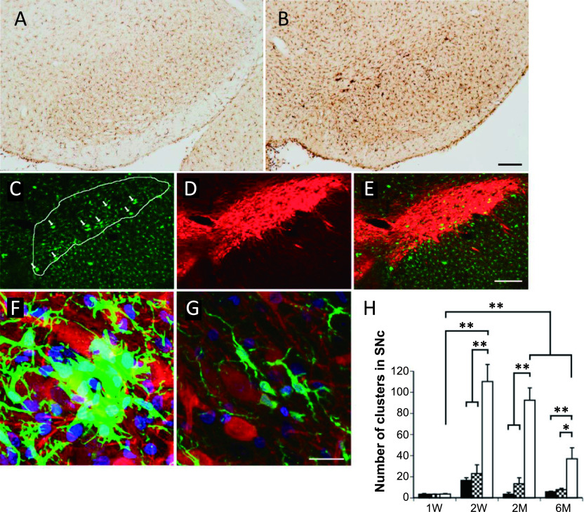
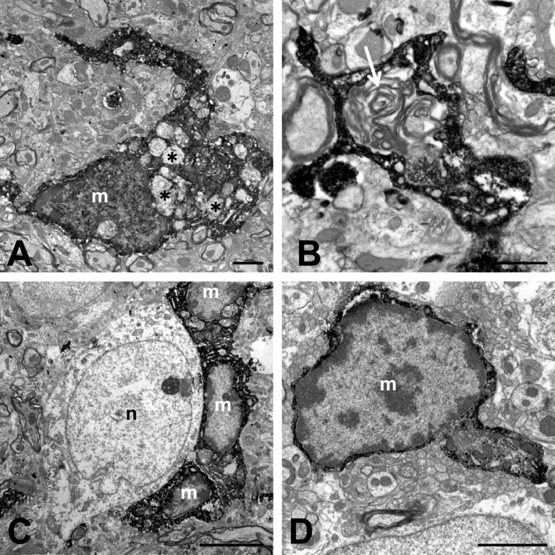
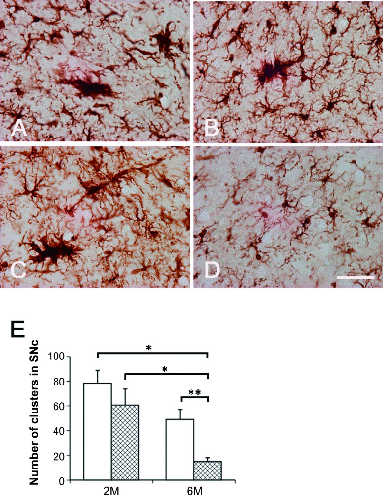
Microglial activation is a component of neurodegenerative pathology. Here, we examine whether activated microglia participate in age-related dopaminergic (DA) cell death in the substantia nigra pars compacta (SNc) of the zitter (zi/zi) rat, a mutant characterized by deletion of the attractin gene. Confocal microscopy with double-immunohistochemical staining revealed activated microglia-formed cell-clusters surrounding DA neurons in the SNc from 2 weeks after birth. An immunoelectron microscopic study showed that the cytoplasm of activated microglia usually contains phagosome-like vacuoles and lamellar inclusions. Expression levels of the pro-inflammatory cytokines interleukin-1β (IL-1β), tumor necrosis factor-α (TNF-α) and inducible nitric oxide synthase (iNOS) were increased in the midbrain of 2-month-old zi/zi rats. Chronic treatment with the anti-inflammatory agent minocycline altered the morphology of the microglia, reduced cluster formation by the microglia, and attenuated DA cell death in the SNc, and reduced the expression of IL-1β in the midbrain. These results indicate that activated microglia, at least in part and especially at the initial phase, contribute to DA cell death in the SNc of the zi/zi rat.
期刊介绍:
Acta Histochemica et Cytochemica is the official online journal of the Japan Society of Histochemistry and Cytochemistry. It is intended primarily for rapid publication of concise, original articles in the fields of histochemistry and cytochemistry. Manuscripts oriented towards methodological subjects that contain significant technical advances in these fields are also welcome. Manuscripts in English are accepted from investigators in any country, whether or not they are members of the Japan Society of Histochemistry and Cytochemistry. Manuscripts should be original work that has not been previously published and is not being considered for publication elsewhere, with the exception of abstracts. Manuscripts with essentially the same content as a paper that has been published or accepted, or is under consideration for publication, will not be considered. All submitted papers will be peer-reviewed by at least two referees selected by an appropriate Associate Editor. Acceptance is based on scientific significance, originality, and clarity. When required, a revised manuscript should be submitted within 3 months, otherwise it will be considered to be a new submission. The Editor-in-Chief will make all final decisions regarding acceptance.
 求助内容:
求助内容: 应助结果提醒方式:


