Isabel Schultz-Pernice, Lisa K Engelbrecht, Stefania Petricca, Christina H Scheel, Alecia-Jane Twigger
{"title":"人乳膜封闭结构的形态学分析揭示了不同的细胞和细胞样乳脂球。","authors":"Isabel Schultz-Pernice, Lisa K Engelbrecht, Stefania Petricca, Christina H Scheel, Alecia-Jane Twigger","doi":"10.1007/s10911-020-09472-1","DOIUrl":null,"url":null,"abstract":"<p><p>Over the past decade, the cellular content of human milk has been a focus in lactation research due to the benefit a potential non-invasive stem cell compartment could provide either to the infant or for therapeutic applications. Despite an increase in the number of studies in this field, fundamental knowledge in regard to milk cell identification and characterisation is still lacking. In this project, we investigated the nature, morphology and content of membrane enclosed structures (MESs) and explored different methods to enrich human milk cells (HMCs) whilst reducing milk fat globule (MFG) content. Using both flow cytometry and immunofluorescence imaging, we confirmed previous reports and showed that nucleated HMCs make up a minority of milk-isolated MESs and are indistinguishable from MFGs without the use of a nuclear stain. HMC heterogeneity was demonstrated by differential uptake of nuclear stains Hoechst 33258 and DRAQ5™ using a novel technique of imaging milk MESs (by embedding them in agar), that enabled examination of both extracellular and intracellular markers. We found that MESs often contain multiple lipid droplets of various sizes and for the first time report that late post-partum human milk contains secretory luminal binucleated cells found across a number of participants. After investigation of different techniques, we found that viably freezing milk cells is an easy and effective method to substantially reduce MFG content of samples. Alternatively, milk MESs can be filtered using a MACS® filter and return a highly viable, though reduced population of milk cells. Using the techniques and findings we've developed in this study; future research may focus on further characterising HMCs and the functional secretory mammary epithelium during lactation.</p>","PeriodicalId":16413,"journal":{"name":"Journal of Mammary Gland Biology and Neoplasia","volume":"25 4","pages":"397-408"},"PeriodicalIF":3.0000,"publicationDate":"2020-12-01","publicationTypes":"Journal Article","fieldsOfStudy":null,"isOpenAccess":false,"openAccessPdf":"https://sci-hub-pdf.com/10.1007/s10911-020-09472-1","citationCount":"4","resultStr":"{\"title\":\"Morphological Analysis of Human Milk Membrane Enclosed Structures Reveals Diverse Cells and Cell-like Milk Fat Globules.\",\"authors\":\"Isabel Schultz-Pernice, Lisa K Engelbrecht, Stefania Petricca, Christina H Scheel, Alecia-Jane Twigger\",\"doi\":\"10.1007/s10911-020-09472-1\",\"DOIUrl\":null,\"url\":null,\"abstract\":\"<p><p>Over the past decade, the cellular content of human milk has been a focus in lactation research due to the benefit a potential non-invasive stem cell compartment could provide either to the infant or for therapeutic applications. Despite an increase in the number of studies in this field, fundamental knowledge in regard to milk cell identification and characterisation is still lacking. In this project, we investigated the nature, morphology and content of membrane enclosed structures (MESs) and explored different methods to enrich human milk cells (HMCs) whilst reducing milk fat globule (MFG) content. Using both flow cytometry and immunofluorescence imaging, we confirmed previous reports and showed that nucleated HMCs make up a minority of milk-isolated MESs and are indistinguishable from MFGs without the use of a nuclear stain. HMC heterogeneity was demonstrated by differential uptake of nuclear stains Hoechst 33258 and DRAQ5™ using a novel technique of imaging milk MESs (by embedding them in agar), that enabled examination of both extracellular and intracellular markers. We found that MESs often contain multiple lipid droplets of various sizes and for the first time report that late post-partum human milk contains secretory luminal binucleated cells found across a number of participants. After investigation of different techniques, we found that viably freezing milk cells is an easy and effective method to substantially reduce MFG content of samples. Alternatively, milk MESs can be filtered using a MACS® filter and return a highly viable, though reduced population of milk cells. Using the techniques and findings we've developed in this study; future research may focus on further characterising HMCs and the functional secretory mammary epithelium during lactation.</p>\",\"PeriodicalId\":16413,\"journal\":{\"name\":\"Journal of Mammary Gland Biology and Neoplasia\",\"volume\":\"25 4\",\"pages\":\"397-408\"},\"PeriodicalIF\":3.0000,\"publicationDate\":\"2020-12-01\",\"publicationTypes\":\"Journal Article\",\"fieldsOfStudy\":null,\"isOpenAccess\":false,\"openAccessPdf\":\"https://sci-hub-pdf.com/10.1007/s10911-020-09472-1\",\"citationCount\":\"4\",\"resultStr\":null,\"platform\":\"Semanticscholar\",\"paperid\":null,\"PeriodicalName\":\"Journal of Mammary Gland Biology and Neoplasia\",\"FirstCategoryId\":\"3\",\"ListUrlMain\":\"https://doi.org/10.1007/s10911-020-09472-1\",\"RegionNum\":4,\"RegionCategory\":\"医学\",\"ArticlePicture\":[],\"TitleCN\":null,\"AbstractTextCN\":null,\"PMCID\":null,\"EPubDate\":\"2021/1/4 0:00:00\",\"PubModel\":\"Epub\",\"JCR\":\"Q2\",\"JCRName\":\"ENDOCRINOLOGY & METABOLISM\",\"Score\":null,\"Total\":0}","platform":"Semanticscholar","paperid":null,"PeriodicalName":"Journal of Mammary Gland Biology and Neoplasia","FirstCategoryId":"3","ListUrlMain":"https://doi.org/10.1007/s10911-020-09472-1","RegionNum":4,"RegionCategory":"医学","ArticlePicture":[],"TitleCN":null,"AbstractTextCN":null,"PMCID":null,"EPubDate":"2021/1/4 0:00:00","PubModel":"Epub","JCR":"Q2","JCRName":"ENDOCRINOLOGY & METABOLISM","Score":null,"Total":0}
Morphological Analysis of Human Milk Membrane Enclosed Structures Reveals Diverse Cells and Cell-like Milk Fat Globules.
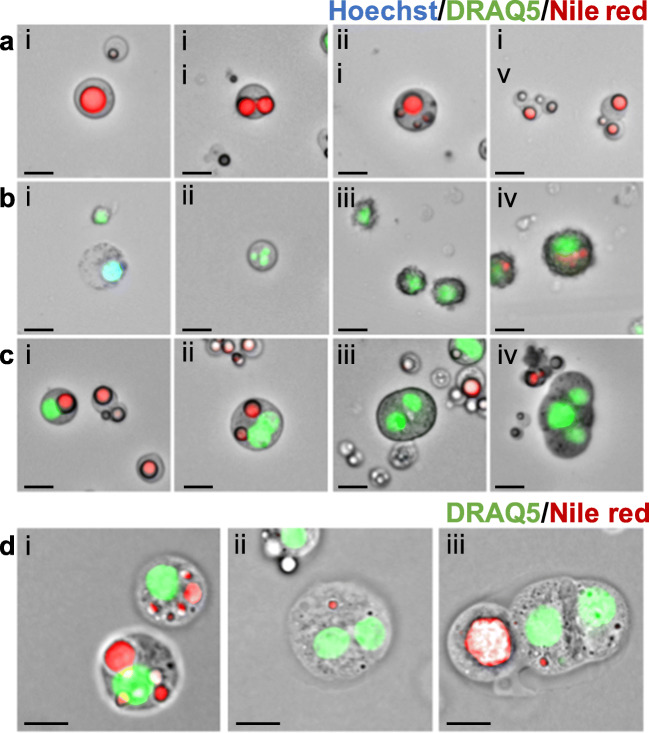
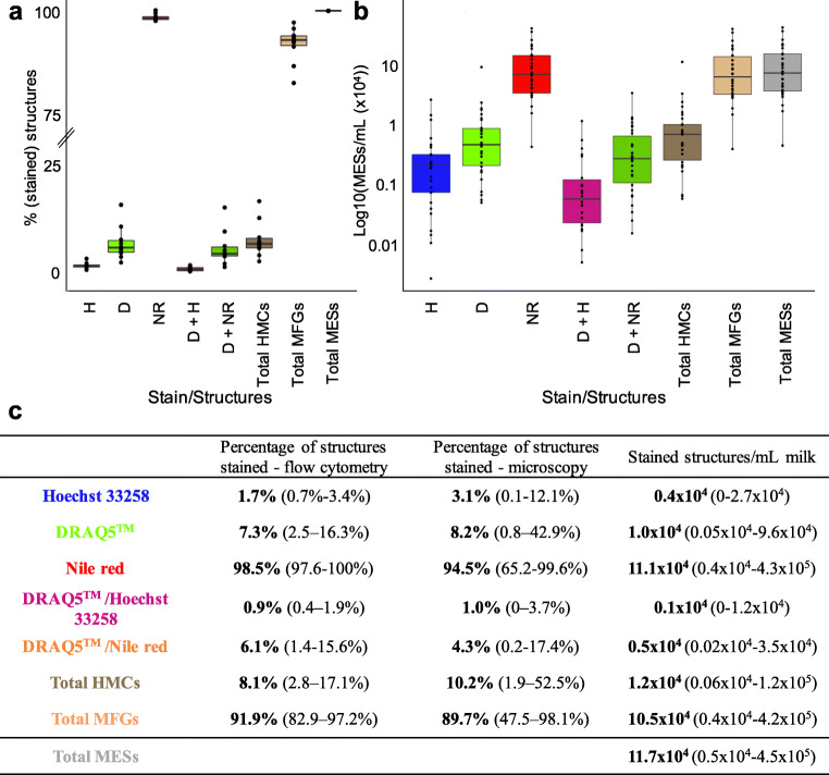
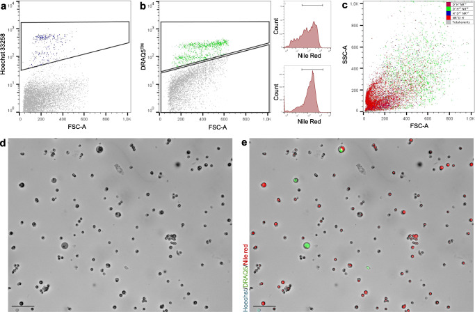
Over the past decade, the cellular content of human milk has been a focus in lactation research due to the benefit a potential non-invasive stem cell compartment could provide either to the infant or for therapeutic applications. Despite an increase in the number of studies in this field, fundamental knowledge in regard to milk cell identification and characterisation is still lacking. In this project, we investigated the nature, morphology and content of membrane enclosed structures (MESs) and explored different methods to enrich human milk cells (HMCs) whilst reducing milk fat globule (MFG) content. Using both flow cytometry and immunofluorescence imaging, we confirmed previous reports and showed that nucleated HMCs make up a minority of milk-isolated MESs and are indistinguishable from MFGs without the use of a nuclear stain. HMC heterogeneity was demonstrated by differential uptake of nuclear stains Hoechst 33258 and DRAQ5™ using a novel technique of imaging milk MESs (by embedding them in agar), that enabled examination of both extracellular and intracellular markers. We found that MESs often contain multiple lipid droplets of various sizes and for the first time report that late post-partum human milk contains secretory luminal binucleated cells found across a number of participants. After investigation of different techniques, we found that viably freezing milk cells is an easy and effective method to substantially reduce MFG content of samples. Alternatively, milk MESs can be filtered using a MACS® filter and return a highly viable, though reduced population of milk cells. Using the techniques and findings we've developed in this study; future research may focus on further characterising HMCs and the functional secretory mammary epithelium during lactation.
期刊介绍:
Journal of Mammary Gland Biology and Neoplasia is the leading Journal in the field of mammary gland biology that provides researchers within and outside the field of mammary gland biology with an integrated source of information pertaining to the development, function, and pathology of the mammary gland and its function.
Commencing in 2015, the Journal will begin receiving and publishing a combination of reviews and original, peer-reviewed research. The Journal covers all topics related to the field of mammary gland biology, including mammary development, breast cancer biology, lactation, and milk composition and quality. The environmental, endocrine, nutritional, and molecular factors regulating these processes is covered, including from a comparative biology perspective.
 求助内容:
求助内容: 应助结果提醒方式:


