Arutselvan Natarajan, Shyam M Srinivas, Carmen Azevedo, Lacey Greene, Anne-Laure Bauchet, Erwan Jouannot, Anne-Sophie Lacoste-Bourgeacq, Isabelle Guizon, Patrick Cohen, Anne-Laure Naneix, Ohad Ilovich, Jordan Cisneros, Krithika Rupanarayan, Frederick T Chin, Andrei Iagaru, Frederick M Dirbas, Amer Karam, Sanjiv S Gambhir
{"title":"两例伴随诊断免疫-正电子发射断层扫描(PET)示踪剂用于检测抗体药物偶联(ADC)治疗中癌症中人类CA6表达的患者研究。","authors":"Arutselvan Natarajan, Shyam M Srinivas, Carmen Azevedo, Lacey Greene, Anne-Laure Bauchet, Erwan Jouannot, Anne-Sophie Lacoste-Bourgeacq, Isabelle Guizon, Patrick Cohen, Anne-Laure Naneix, Ohad Ilovich, Jordan Cisneros, Krithika Rupanarayan, Frederick T Chin, Andrei Iagaru, Frederick M Dirbas, Amer Karam, Sanjiv S Gambhir","doi":"10.1177/1536012120939398","DOIUrl":null,"url":null,"abstract":"<p><p>An antigen binding fragment (BFab) derived from a tumor-associated mucin 1-sialoglycotope antigen (CA6) targeting antibody (huDS6) was engineered. We synthesized a companion diagnostic positron emission tomography (PET) tracer by radiolabeling BFab with [<sup>64</sup>Cu] to measure CA6 expression on cancer tissues prior to anti-human CA6 (huDS6-DM4 antibody-drug conjugate) therapy for ovarian and breast cancer patients. After chemotherapy, the ovarian patient received PET scan with <sup>18</sup>F-2-fluoro-2-deoxyglucose ([<sup>18</sup>F]FDG: 10 mCi), followed by [<sup>64</sup>Cu]-DOTA-BFab ([<sup>64</sup>Cu]BFab; 5.5 mCi) 1 week later for PET scanning of CA6 expression and subsequent surgery. The breast cancer patient was treated with chemotherapy before primary tumor resection and subsequent [<sup>18</sup>F]FDG-PET scan. 4 weeks later the patient received of [<sup>64</sup>Cu]BFab (11.7 mCi) for CA6 PET scan. Whole body [<sup>18</sup>F]FDG-PET of the breast cancer patient indicated FDG-avid tumor metastases to the liver, bilateral hila and thoracic spine, but no uptake was observed for the ovarian patient. Each patient was also imaged by PET/CT with [<sup>64</sup>Cu]BFab at 1 and 24 hours after tracer administration. The [<sup>64</sup>Cu]BFab tracer was well tolerated by both patients without adverse effects, and no significant tracer uptake was observed in both patients. Immunohistochemistry (IHC) data indicated CA6 expressions were weak to intermediate and matched with the [<sup>64</sup>Cu]BFab-PET signals.</p>","PeriodicalId":18855,"journal":{"name":"Molecular Imaging","volume":"19 ","pages":"1536012120939398"},"PeriodicalIF":2.4000,"publicationDate":"2020-01-01","publicationTypes":"Journal Article","fieldsOfStudy":null,"isOpenAccess":false,"openAccessPdf":"https://sci-hub-pdf.com/10.1177/1536012120939398","citationCount":"4","resultStr":"{\"title\":\"Two Patient Studies of a Companion Diagnostic Immuno-Positron Emission Tomography (PET) Tracer for Measuring Human CA6 Expression in Cancer for Antibody Drug Conjugate (ADC) Therapy.\",\"authors\":\"Arutselvan Natarajan, Shyam M Srinivas, Carmen Azevedo, Lacey Greene, Anne-Laure Bauchet, Erwan Jouannot, Anne-Sophie Lacoste-Bourgeacq, Isabelle Guizon, Patrick Cohen, Anne-Laure Naneix, Ohad Ilovich, Jordan Cisneros, Krithika Rupanarayan, Frederick T Chin, Andrei Iagaru, Frederick M Dirbas, Amer Karam, Sanjiv S Gambhir\",\"doi\":\"10.1177/1536012120939398\",\"DOIUrl\":null,\"url\":null,\"abstract\":\"<p><p>An antigen binding fragment (BFab) derived from a tumor-associated mucin 1-sialoglycotope antigen (CA6) targeting antibody (huDS6) was engineered. We synthesized a companion diagnostic positron emission tomography (PET) tracer by radiolabeling BFab with [<sup>64</sup>Cu] to measure CA6 expression on cancer tissues prior to anti-human CA6 (huDS6-DM4 antibody-drug conjugate) therapy for ovarian and breast cancer patients. After chemotherapy, the ovarian patient received PET scan with <sup>18</sup>F-2-fluoro-2-deoxyglucose ([<sup>18</sup>F]FDG: 10 mCi), followed by [<sup>64</sup>Cu]-DOTA-BFab ([<sup>64</sup>Cu]BFab; 5.5 mCi) 1 week later for PET scanning of CA6 expression and subsequent surgery. The breast cancer patient was treated with chemotherapy before primary tumor resection and subsequent [<sup>18</sup>F]FDG-PET scan. 4 weeks later the patient received of [<sup>64</sup>Cu]BFab (11.7 mCi) for CA6 PET scan. Whole body [<sup>18</sup>F]FDG-PET of the breast cancer patient indicated FDG-avid tumor metastases to the liver, bilateral hila and thoracic spine, but no uptake was observed for the ovarian patient. Each patient was also imaged by PET/CT with [<sup>64</sup>Cu]BFab at 1 and 24 hours after tracer administration. The [<sup>64</sup>Cu]BFab tracer was well tolerated by both patients without adverse effects, and no significant tracer uptake was observed in both patients. Immunohistochemistry (IHC) data indicated CA6 expressions were weak to intermediate and matched with the [<sup>64</sup>Cu]BFab-PET signals.</p>\",\"PeriodicalId\":18855,\"journal\":{\"name\":\"Molecular Imaging\",\"volume\":\"19 \",\"pages\":\"1536012120939398\"},\"PeriodicalIF\":2.4000,\"publicationDate\":\"2020-01-01\",\"publicationTypes\":\"Journal Article\",\"fieldsOfStudy\":null,\"isOpenAccess\":false,\"openAccessPdf\":\"https://sci-hub-pdf.com/10.1177/1536012120939398\",\"citationCount\":\"4\",\"resultStr\":null,\"platform\":\"Semanticscholar\",\"paperid\":null,\"PeriodicalName\":\"Molecular Imaging\",\"FirstCategoryId\":\"3\",\"ListUrlMain\":\"https://doi.org/10.1177/1536012120939398\",\"RegionNum\":4,\"RegionCategory\":\"医学\",\"ArticlePicture\":[],\"TitleCN\":null,\"AbstractTextCN\":null,\"PMCID\":null,\"EPubDate\":\"\",\"PubModel\":\"\",\"JCR\":\"Q3\",\"JCRName\":\"BIOCHEMICAL RESEARCH METHODS\",\"Score\":null,\"Total\":0}","platform":"Semanticscholar","paperid":null,"PeriodicalName":"Molecular Imaging","FirstCategoryId":"3","ListUrlMain":"https://doi.org/10.1177/1536012120939398","RegionNum":4,"RegionCategory":"医学","ArticlePicture":[],"TitleCN":null,"AbstractTextCN":null,"PMCID":null,"EPubDate":"","PubModel":"","JCR":"Q3","JCRName":"BIOCHEMICAL RESEARCH METHODS","Score":null,"Total":0}
Two Patient Studies of a Companion Diagnostic Immuno-Positron Emission Tomography (PET) Tracer for Measuring Human CA6 Expression in Cancer for Antibody Drug Conjugate (ADC) Therapy.
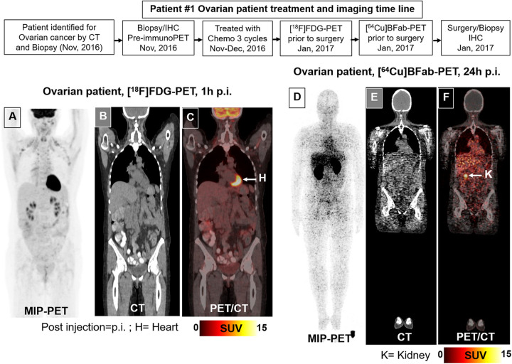
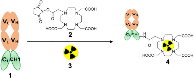
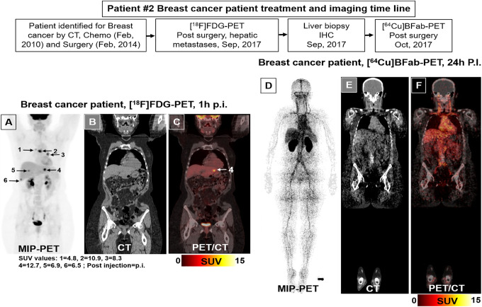
An antigen binding fragment (BFab) derived from a tumor-associated mucin 1-sialoglycotope antigen (CA6) targeting antibody (huDS6) was engineered. We synthesized a companion diagnostic positron emission tomography (PET) tracer by radiolabeling BFab with [64Cu] to measure CA6 expression on cancer tissues prior to anti-human CA6 (huDS6-DM4 antibody-drug conjugate) therapy for ovarian and breast cancer patients. After chemotherapy, the ovarian patient received PET scan with 18F-2-fluoro-2-deoxyglucose ([18F]FDG: 10 mCi), followed by [64Cu]-DOTA-BFab ([64Cu]BFab; 5.5 mCi) 1 week later for PET scanning of CA6 expression and subsequent surgery. The breast cancer patient was treated with chemotherapy before primary tumor resection and subsequent [18F]FDG-PET scan. 4 weeks later the patient received of [64Cu]BFab (11.7 mCi) for CA6 PET scan. Whole body [18F]FDG-PET of the breast cancer patient indicated FDG-avid tumor metastases to the liver, bilateral hila and thoracic spine, but no uptake was observed for the ovarian patient. Each patient was also imaged by PET/CT with [64Cu]BFab at 1 and 24 hours after tracer administration. The [64Cu]BFab tracer was well tolerated by both patients without adverse effects, and no significant tracer uptake was observed in both patients. Immunohistochemistry (IHC) data indicated CA6 expressions were weak to intermediate and matched with the [64Cu]BFab-PET signals.
Molecular ImagingBiochemistry, Genetics and Molecular Biology-Biotechnology
自引率
3.60%
发文量
21
期刊介绍:
Molecular Imaging is a peer-reviewed, open access journal highlighting the breadth of molecular imaging research from basic science to preclinical studies to human applications. This serves both the scientific and clinical communities by disseminating novel results and concepts relevant to the biological study of normal and disease processes in both basic and translational studies ranging from mice to humans.
 求助内容:
求助内容: 应助结果提醒方式:


