{"title":"一种新的抗菌策略:组蛋白和抗菌肽协同作用。","authors":"Leora Duong, Steven P Gross, Albert Siryaporn","doi":"10.15698/mic2020.11.736","DOIUrl":null,"url":null,"abstract":"<p><p>The rate at which antibiotics are discovered and developed has stagnated; meanwhile, antibacterial resistance continually increases and leads to a plethora of untreatable and deadly infections worldwide. Therefore, there is a critical need to develop new antimicrobial strategies to combat this alarming reality. One approach is to understand natural antimicrobial defense mechanisms that higher-level organisms employ in order to kill bacteria, potentially leading to novel antibiotic therapeutic approaches. Mammalian histones have long been reported to have antibiotic activity, with the first observation of their antibacterial properties reported in 1942. However, there have been doubts about whether histones could truly have any such role in the animal, predominantly based on two issues: they are found in the nucleus (so are not in a position to encounter bacteria), and their antibiotic activity <i>in vitro</i> has been relatively weak in physiological conditions. More recent studies have addressed both sets of concerns. Histones are released from cells as part of neutrophil extracellular traps (NETs) and are thus able to encounter extracellular bacteria. Histones are also present intracellularly in the cytoplasm attached to lipid droplets, positioning them to encounter cytosolic bacteria. Our recent work (Doolin et al., 2020, Nat Commun), which is discussed here, shows that histones have synergistic antimicrobial activities when they are paired with antimicrobial peptides (AMPs), which form pores in bacterial membranes and co-localize with histones in NETs. The work demonstrates that histones enhance AMP-mediated pores, impair bacterial membrane recovery, depolarize the bacterial proton gradient, and enter the bacterial cytoplasm, where they restructure the chromosome and inhibit transcription. Here, we examine potential mechanisms that are responsible for these outcomes.</p>","PeriodicalId":18397,"journal":{"name":"Microbial Cell","volume":"7 11","pages":"309-311"},"PeriodicalIF":3.9000,"publicationDate":"2020-10-08","publicationTypes":"Journal Article","fieldsOfStudy":null,"isOpenAccess":false,"openAccessPdf":"https://www.ncbi.nlm.nih.gov/pmc/articles/PMC7590529/pdf/","citationCount":"6","resultStr":"{\"title\":\"A novel antibacterial strategy: histone and antimicrobial peptide synergy.\",\"authors\":\"Leora Duong, Steven P Gross, Albert Siryaporn\",\"doi\":\"10.15698/mic2020.11.736\",\"DOIUrl\":null,\"url\":null,\"abstract\":\"<p><p>The rate at which antibiotics are discovered and developed has stagnated; meanwhile, antibacterial resistance continually increases and leads to a plethora of untreatable and deadly infections worldwide. Therefore, there is a critical need to develop new antimicrobial strategies to combat this alarming reality. One approach is to understand natural antimicrobial defense mechanisms that higher-level organisms employ in order to kill bacteria, potentially leading to novel antibiotic therapeutic approaches. Mammalian histones have long been reported to have antibiotic activity, with the first observation of their antibacterial properties reported in 1942. However, there have been doubts about whether histones could truly have any such role in the animal, predominantly based on two issues: they are found in the nucleus (so are not in a position to encounter bacteria), and their antibiotic activity <i>in vitro</i> has been relatively weak in physiological conditions. More recent studies have addressed both sets of concerns. Histones are released from cells as part of neutrophil extracellular traps (NETs) and are thus able to encounter extracellular bacteria. Histones are also present intracellularly in the cytoplasm attached to lipid droplets, positioning them to encounter cytosolic bacteria. Our recent work (Doolin et al., 2020, Nat Commun), which is discussed here, shows that histones have synergistic antimicrobial activities when they are paired with antimicrobial peptides (AMPs), which form pores in bacterial membranes and co-localize with histones in NETs. The work demonstrates that histones enhance AMP-mediated pores, impair bacterial membrane recovery, depolarize the bacterial proton gradient, and enter the bacterial cytoplasm, where they restructure the chromosome and inhibit transcription. Here, we examine potential mechanisms that are responsible for these outcomes.</p>\",\"PeriodicalId\":18397,\"journal\":{\"name\":\"Microbial Cell\",\"volume\":\"7 11\",\"pages\":\"309-311\"},\"PeriodicalIF\":3.9000,\"publicationDate\":\"2020-10-08\",\"publicationTypes\":\"Journal Article\",\"fieldsOfStudy\":null,\"isOpenAccess\":false,\"openAccessPdf\":\"https://www.ncbi.nlm.nih.gov/pmc/articles/PMC7590529/pdf/\",\"citationCount\":\"6\",\"resultStr\":null,\"platform\":\"Semanticscholar\",\"paperid\":null,\"PeriodicalName\":\"Microbial Cell\",\"FirstCategoryId\":\"99\",\"ListUrlMain\":\"https://doi.org/10.15698/mic2020.11.736\",\"RegionNum\":3,\"RegionCategory\":\"生物学\",\"ArticlePicture\":[],\"TitleCN\":null,\"AbstractTextCN\":null,\"PMCID\":null,\"EPubDate\":\"\",\"PubModel\":\"\",\"JCR\":\"Q2\",\"JCRName\":\"CELL BIOLOGY\",\"Score\":null,\"Total\":0}","platform":"Semanticscholar","paperid":null,"PeriodicalName":"Microbial Cell","FirstCategoryId":"99","ListUrlMain":"https://doi.org/10.15698/mic2020.11.736","RegionNum":3,"RegionCategory":"生物学","ArticlePicture":[],"TitleCN":null,"AbstractTextCN":null,"PMCID":null,"EPubDate":"","PubModel":"","JCR":"Q2","JCRName":"CELL BIOLOGY","Score":null,"Total":0}
引用次数: 6
摘要
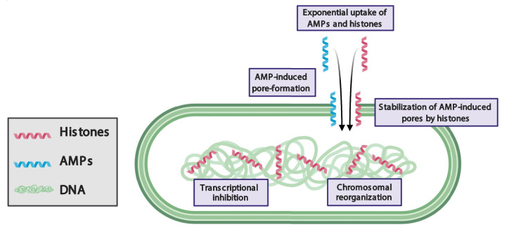
抗生素的发现和开发速度停滞不前;与此同时,抗菌素耐药性不断增加,导致世界范围内出现大量无法治疗和致命的感染。因此,迫切需要制定新的抗微生物战略来应对这一令人震惊的现实。一种方法是了解高级生物为了杀死细菌而采用的天然抗菌防御机制,这可能会导致新的抗生素治疗方法。哺乳动物组蛋白长期以来一直被报道具有抗生素活性,1942年首次观察到它们的抗菌特性。然而,对于组蛋白在动物体内是否真的具有这样的作用一直存在怀疑,主要基于两个问题:组蛋白存在于细胞核中(因此无法与细菌接触),以及它们在体外的抗生素活性在生理条件下相对较弱。最近的研究解决了这两种问题。组蛋白作为中性粒细胞胞外陷阱(NETs)的一部分从细胞中释放出来,因此能够遇到胞外细菌。组蛋白也存在于细胞内的细胞质中,附着在脂滴上,使它们能够遇到胞质细菌。我们最近的研究(Doolin et al., 2020, Nat comm)表明,当组蛋白与抗菌肽(抗菌肽在细菌膜上形成孔,并在NETs中与组蛋白共定位)结合时,组蛋白具有协同抗菌活性。研究表明,组蛋白增强amp介导的孔隙,损害细菌膜恢复,使细菌质子梯度去极化,并进入细菌细胞质,在那里它们重组染色体并抑制转录。在这里,我们研究了导致这些结果的潜在机制。
A novel antibacterial strategy: histone and antimicrobial peptide synergy.
The rate at which antibiotics are discovered and developed has stagnated; meanwhile, antibacterial resistance continually increases and leads to a plethora of untreatable and deadly infections worldwide. Therefore, there is a critical need to develop new antimicrobial strategies to combat this alarming reality. One approach is to understand natural antimicrobial defense mechanisms that higher-level organisms employ in order to kill bacteria, potentially leading to novel antibiotic therapeutic approaches. Mammalian histones have long been reported to have antibiotic activity, with the first observation of their antibacterial properties reported in 1942. However, there have been doubts about whether histones could truly have any such role in the animal, predominantly based on two issues: they are found in the nucleus (so are not in a position to encounter bacteria), and their antibiotic activity in vitro has been relatively weak in physiological conditions. More recent studies have addressed both sets of concerns. Histones are released from cells as part of neutrophil extracellular traps (NETs) and are thus able to encounter extracellular bacteria. Histones are also present intracellularly in the cytoplasm attached to lipid droplets, positioning them to encounter cytosolic bacteria. Our recent work (Doolin et al., 2020, Nat Commun), which is discussed here, shows that histones have synergistic antimicrobial activities when they are paired with antimicrobial peptides (AMPs), which form pores in bacterial membranes and co-localize with histones in NETs. The work demonstrates that histones enhance AMP-mediated pores, impair bacterial membrane recovery, depolarize the bacterial proton gradient, and enter the bacterial cytoplasm, where they restructure the chromosome and inhibit transcription. Here, we examine potential mechanisms that are responsible for these outcomes.

 求助内容:
求助内容: 应助结果提醒方式:


