Sukritee Bhaskar , David L. Steer , Ruchi Anand , Santosh Panjikar
{"title":"区分两类硫醇酶的结构基础:降解型与生物合成型硫醇酶","authors":"Sukritee Bhaskar , David L. Steer , Ruchi Anand , Santosh Panjikar","doi":"10.1016/j.yjsbx.2019.100018","DOIUrl":null,"url":null,"abstract":"<div><p>Thiolases are a well characterized family of enzymes with two distinct categories: degradative, β-ketoadipyl-CoA thiolases and biosynthetic, acetoacetyl-CoA thiolases. Both classes share an identical catalytic triad but catalyze reactions in opposite directions. Moreover, it is established that in contrast to the biosynthetic thiolases the degradative thiolases can accept substrates with broad chain lengths. Hitherto, no residue or structural pattern has been recognized that might help to discern the two thiolases, here we exploit, a tetrameric degradative thiolase from <em>Pseudomonas putida</em> KT2440 annotated as PcaF, as a model system to understand features which distinguishes the two classes using structural studies and bioinformatics analyses. Degradative thiolases have different active site architecture when compared to biosynthetic thiolases, demonstrating the dissimilar chemical nature of the active site architecture. Both thiolases deploy different “anchoring residues” to tether the large Coenzyme A (CoA) or CoA derivatives. Interestingly, the H356 of the catalytic triad in PcaF is directly involved in tethering the CoA/CoA derivatives into the active site and we were able to trap a gridlocked thiolase structure of the H356A mutant, where the CoA was found to be covalently linked to the catalytic cysteine residue, inhibiting the overall reaction. Further, X-ray structures with two long chain CoA derivatives, hexanal-CoA and octanal-CoA helped in delineating the long tunnel of 235 Å<sup>2</sup> surface area in PcaF and led to identification of a unique covering loop exclusive to degradative thiolases that plays an active role in determining the tunnel length and the nature of the binding substrate.</p></div>","PeriodicalId":17238,"journal":{"name":"Journal of Structural Biology: X","volume":"4 ","pages":"Article 100018"},"PeriodicalIF":3.5000,"publicationDate":"2020-01-01","publicationTypes":"Journal Article","fieldsOfStudy":null,"isOpenAccess":false,"openAccessPdf":"https://sci-hub-pdf.com/10.1016/j.yjsbx.2019.100018","citationCount":"7","resultStr":"{\"title\":\"Structural basis for differentiation between two classes of thiolase: Degradative vs biosynthetic thiolase\",\"authors\":\"Sukritee Bhaskar , David L. Steer , Ruchi Anand , Santosh Panjikar\",\"doi\":\"10.1016/j.yjsbx.2019.100018\",\"DOIUrl\":null,\"url\":null,\"abstract\":\"<div><p>Thiolases are a well characterized family of enzymes with two distinct categories: degradative, β-ketoadipyl-CoA thiolases and biosynthetic, acetoacetyl-CoA thiolases. Both classes share an identical catalytic triad but catalyze reactions in opposite directions. Moreover, it is established that in contrast to the biosynthetic thiolases the degradative thiolases can accept substrates with broad chain lengths. Hitherto, no residue or structural pattern has been recognized that might help to discern the two thiolases, here we exploit, a tetrameric degradative thiolase from <em>Pseudomonas putida</em> KT2440 annotated as PcaF, as a model system to understand features which distinguishes the two classes using structural studies and bioinformatics analyses. Degradative thiolases have different active site architecture when compared to biosynthetic thiolases, demonstrating the dissimilar chemical nature of the active site architecture. Both thiolases deploy different “anchoring residues” to tether the large Coenzyme A (CoA) or CoA derivatives. Interestingly, the H356 of the catalytic triad in PcaF is directly involved in tethering the CoA/CoA derivatives into the active site and we were able to trap a gridlocked thiolase structure of the H356A mutant, where the CoA was found to be covalently linked to the catalytic cysteine residue, inhibiting the overall reaction. Further, X-ray structures with two long chain CoA derivatives, hexanal-CoA and octanal-CoA helped in delineating the long tunnel of 235 Å<sup>2</sup> surface area in PcaF and led to identification of a unique covering loop exclusive to degradative thiolases that plays an active role in determining the tunnel length and the nature of the binding substrate.</p></div>\",\"PeriodicalId\":17238,\"journal\":{\"name\":\"Journal of Structural Biology: X\",\"volume\":\"4 \",\"pages\":\"Article 100018\"},\"PeriodicalIF\":3.5000,\"publicationDate\":\"2020-01-01\",\"publicationTypes\":\"Journal Article\",\"fieldsOfStudy\":null,\"isOpenAccess\":false,\"openAccessPdf\":\"https://sci-hub-pdf.com/10.1016/j.yjsbx.2019.100018\",\"citationCount\":\"7\",\"resultStr\":null,\"platform\":\"Semanticscholar\",\"paperid\":null,\"PeriodicalName\":\"Journal of Structural Biology: X\",\"FirstCategoryId\":\"1085\",\"ListUrlMain\":\"https://www.sciencedirect.com/science/article/pii/S2590152419300169\",\"RegionNum\":0,\"RegionCategory\":null,\"ArticlePicture\":[],\"TitleCN\":null,\"AbstractTextCN\":null,\"PMCID\":null,\"EPubDate\":\"\",\"PubModel\":\"\",\"JCR\":\"Q2\",\"JCRName\":\"BIOCHEMISTRY & MOLECULAR BIOLOGY\",\"Score\":null,\"Total\":0}","platform":"Semanticscholar","paperid":null,"PeriodicalName":"Journal of Structural Biology: X","FirstCategoryId":"1085","ListUrlMain":"https://www.sciencedirect.com/science/article/pii/S2590152419300169","RegionNum":0,"RegionCategory":null,"ArticlePicture":[],"TitleCN":null,"AbstractTextCN":null,"PMCID":null,"EPubDate":"","PubModel":"","JCR":"Q2","JCRName":"BIOCHEMISTRY & MOLECULAR BIOLOGY","Score":null,"Total":0}
Structural basis for differentiation between two classes of thiolase: Degradative vs biosynthetic thiolase
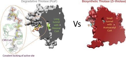
Thiolases are a well characterized family of enzymes with two distinct categories: degradative, β-ketoadipyl-CoA thiolases and biosynthetic, acetoacetyl-CoA thiolases. Both classes share an identical catalytic triad but catalyze reactions in opposite directions. Moreover, it is established that in contrast to the biosynthetic thiolases the degradative thiolases can accept substrates with broad chain lengths. Hitherto, no residue or structural pattern has been recognized that might help to discern the two thiolases, here we exploit, a tetrameric degradative thiolase from Pseudomonas putida KT2440 annotated as PcaF, as a model system to understand features which distinguishes the two classes using structural studies and bioinformatics analyses. Degradative thiolases have different active site architecture when compared to biosynthetic thiolases, demonstrating the dissimilar chemical nature of the active site architecture. Both thiolases deploy different “anchoring residues” to tether the large Coenzyme A (CoA) or CoA derivatives. Interestingly, the H356 of the catalytic triad in PcaF is directly involved in tethering the CoA/CoA derivatives into the active site and we were able to trap a gridlocked thiolase structure of the H356A mutant, where the CoA was found to be covalently linked to the catalytic cysteine residue, inhibiting the overall reaction. Further, X-ray structures with two long chain CoA derivatives, hexanal-CoA and octanal-CoA helped in delineating the long tunnel of 235 Å2 surface area in PcaF and led to identification of a unique covering loop exclusive to degradative thiolases that plays an active role in determining the tunnel length and the nature of the binding substrate.
 求助内容:
求助内容: 应助结果提醒方式:


