Nithya Ramakrishnan, Nicholas R E Murphy, Christopher P Walker, Valeria A Cuellar Leal, Jair C Soares, Raymond Y J Cho, Sudhakar Selvaraj
{"title":"氯胺酮对难治性抑郁症患者前额叶皮质的神经生理影响:经颅磁刺激-脑电图联合研究。","authors":"Nithya Ramakrishnan, Nicholas R E Murphy, Christopher P Walker, Valeria A Cuellar Leal, Jair C Soares, Raymond Y J Cho, Sudhakar Selvaraj","doi":"10.1177/2470547019861417","DOIUrl":null,"url":null,"abstract":"Treatment-resistant depression (TRD) represents a substantial clinical and economic burden. A single subanesthetic dose of the noncompetitive N-methyl-D-aspartate (NMDA) receptor antagonist ketamine improves TRD depression symptoms within hours. The rapid response points to a fundamentally different mechanism which, while well modeled in preclinical studies, has yet to be translated into clinically relevant biomarkers. Transcranial magnetic stimulation (TMS)-evoked potentials (TEPs) are a direct index of the neurophysiological state of the stimulated cortical and cortico-thalamic network. TEPs have also previously shown a relationship with glutamatergic and Y-amino butyric acid (GABA)ergic neurotransmission suggesting that concurrent TMS–electroencephalography (EEG) can also be an index of local cortical excitability/inhibition balance. Animal studies suggest that ketamine not only increases glutamatergic excitatory drive in the prefrontal cortex (PFC) and limbic regions of the brain but also demonstrates GABAAR agonism. 3 This study aimed to observe changes in PFC cortical excitability measures indexed by a pharmaco-TMS–EEG approach by evaluating alterations in its component structure up to 24 hours postketamine infusion. Four TRD patients (mean age: 38.3 10.6 years; N 1⁄4 three females) provided written informed consent to participate. They received open-label intravenous infusion of 0.5mg/kg ketamine over 40 minutes. Patient’s depression levels were assessed using the Montgomery–Åsberg Depression Rating Scale (MADRS) and the Hamilton Depression Rating Scale (HAM-D) at pre-ketamine baseline, and 4 hours and 24 hours post-ketamine infusion. Concurrent TMS stimulation and EEG recording were performed at all sessions. Biphasic single-pulse TMS (MagVenture MagPro) was presented at the left dorsolateral prefrontal cortex (DLPFC), for N1⁄4 200 pulses. The cortical response to TMS was recorded using 64-channel EEG (BrainAmp DC, BrainProducts), sampled at 5000Hz, with electrode wires reoriented to avoid direct contact with the TMS coil. Stimulation intensity was 120% of baseline resting motor threshold. EEG data were analyzed by replacing the TMS pulse period (0–20ms) with linear interpolation. Artifacts were removed using a two-tiered independent components analysis routine (ARTIST). This algorithm automatically identifies artifactual components based on features capturing the spatiotemporal profile of both neural and artifactual activities. Additional noise suppression employed the source-estimate-utilizing noisediscarding algorithm (SOUND). We utilized the local mean field amplitude–area under the curve (LMFA– AUC) from a subset of electrodes (Figure 1(d)) around the stimulation site as our primary outcome measure. This has previously been reported as a reliable index of cortical reactivity or excitation. We applied the SOUND correction to individual trials to test within-subject differences from session to session using nonparametric Kruskal–Wallis tests. Overall, patients showed a reduction in the LMFA–AUC at 4 hours that increased back at 24 hours but remained lower than baseline. We also found an overall reduction in peak-to-peak measures at","PeriodicalId":52315,"journal":{"name":"Chronic Stress","volume":" ","pages":"2470547019861417"},"PeriodicalIF":0.0000,"publicationDate":"2019-07-23","publicationTypes":"Journal Article","fieldsOfStudy":null,"isOpenAccess":false,"openAccessPdf":"https://sci-hub-pdf.com/10.1177/2470547019861417","citationCount":"3","resultStr":"{\"title\":\"Neurophysiological Effect of Ketamine on Prefrontal Cortex in Treatment-Resistant Depression: A Combined Transcranial Magnetic Stimulation-Electroencephalography Study.\",\"authors\":\"Nithya Ramakrishnan, Nicholas R E Murphy, Christopher P Walker, Valeria A Cuellar Leal, Jair C Soares, Raymond Y J Cho, Sudhakar Selvaraj\",\"doi\":\"10.1177/2470547019861417\",\"DOIUrl\":null,\"url\":null,\"abstract\":\"Treatment-resistant depression (TRD) represents a substantial clinical and economic burden. A single subanesthetic dose of the noncompetitive N-methyl-D-aspartate (NMDA) receptor antagonist ketamine improves TRD depression symptoms within hours. The rapid response points to a fundamentally different mechanism which, while well modeled in preclinical studies, has yet to be translated into clinically relevant biomarkers. Transcranial magnetic stimulation (TMS)-evoked potentials (TEPs) are a direct index of the neurophysiological state of the stimulated cortical and cortico-thalamic network. TEPs have also previously shown a relationship with glutamatergic and Y-amino butyric acid (GABA)ergic neurotransmission suggesting that concurrent TMS–electroencephalography (EEG) can also be an index of local cortical excitability/inhibition balance. Animal studies suggest that ketamine not only increases glutamatergic excitatory drive in the prefrontal cortex (PFC) and limbic regions of the brain but also demonstrates GABAAR agonism. 3 This study aimed to observe changes in PFC cortical excitability measures indexed by a pharmaco-TMS–EEG approach by evaluating alterations in its component structure up to 24 hours postketamine infusion. Four TRD patients (mean age: 38.3 10.6 years; N 1⁄4 three females) provided written informed consent to participate. They received open-label intravenous infusion of 0.5mg/kg ketamine over 40 minutes. Patient’s depression levels were assessed using the Montgomery–Åsberg Depression Rating Scale (MADRS) and the Hamilton Depression Rating Scale (HAM-D) at pre-ketamine baseline, and 4 hours and 24 hours post-ketamine infusion. Concurrent TMS stimulation and EEG recording were performed at all sessions. Biphasic single-pulse TMS (MagVenture MagPro) was presented at the left dorsolateral prefrontal cortex (DLPFC), for N1⁄4 200 pulses. The cortical response to TMS was recorded using 64-channel EEG (BrainAmp DC, BrainProducts), sampled at 5000Hz, with electrode wires reoriented to avoid direct contact with the TMS coil. Stimulation intensity was 120% of baseline resting motor threshold. EEG data were analyzed by replacing the TMS pulse period (0–20ms) with linear interpolation. Artifacts were removed using a two-tiered independent components analysis routine (ARTIST). This algorithm automatically identifies artifactual components based on features capturing the spatiotemporal profile of both neural and artifactual activities. Additional noise suppression employed the source-estimate-utilizing noisediscarding algorithm (SOUND). We utilized the local mean field amplitude–area under the curve (LMFA– AUC) from a subset of electrodes (Figure 1(d)) around the stimulation site as our primary outcome measure. This has previously been reported as a reliable index of cortical reactivity or excitation. We applied the SOUND correction to individual trials to test within-subject differences from session to session using nonparametric Kruskal–Wallis tests. Overall, patients showed a reduction in the LMFA–AUC at 4 hours that increased back at 24 hours but remained lower than baseline. We also found an overall reduction in peak-to-peak measures at\",\"PeriodicalId\":52315,\"journal\":{\"name\":\"Chronic Stress\",\"volume\":\" \",\"pages\":\"2470547019861417\"},\"PeriodicalIF\":0.0000,\"publicationDate\":\"2019-07-23\",\"publicationTypes\":\"Journal Article\",\"fieldsOfStudy\":null,\"isOpenAccess\":false,\"openAccessPdf\":\"https://sci-hub-pdf.com/10.1177/2470547019861417\",\"citationCount\":\"3\",\"resultStr\":null,\"platform\":\"Semanticscholar\",\"paperid\":null,\"PeriodicalName\":\"Chronic Stress\",\"FirstCategoryId\":\"1085\",\"ListUrlMain\":\"https://doi.org/10.1177/2470547019861417\",\"RegionNum\":0,\"RegionCategory\":null,\"ArticlePicture\":[],\"TitleCN\":null,\"AbstractTextCN\":null,\"PMCID\":null,\"EPubDate\":\"2019/1/1 0:00:00\",\"PubModel\":\"eCollection\",\"JCR\":\"Q1\",\"JCRName\":\"Psychology\",\"Score\":null,\"Total\":0}","platform":"Semanticscholar","paperid":null,"PeriodicalName":"Chronic Stress","FirstCategoryId":"1085","ListUrlMain":"https://doi.org/10.1177/2470547019861417","RegionNum":0,"RegionCategory":null,"ArticlePicture":[],"TitleCN":null,"AbstractTextCN":null,"PMCID":null,"EPubDate":"2019/1/1 0:00:00","PubModel":"eCollection","JCR":"Q1","JCRName":"Psychology","Score":null,"Total":0}
Neurophysiological Effect of Ketamine on Prefrontal Cortex in Treatment-Resistant Depression: A Combined Transcranial Magnetic Stimulation-Electroencephalography Study.
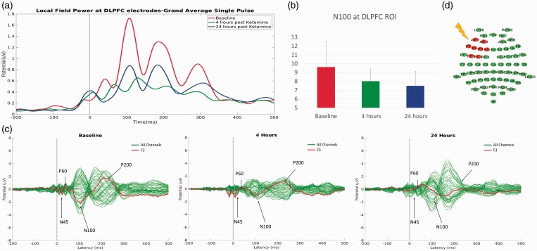
Treatment-resistant depression (TRD) represents a substantial clinical and economic burden. A single subanesthetic dose of the noncompetitive N-methyl-D-aspartate (NMDA) receptor antagonist ketamine improves TRD depression symptoms within hours. The rapid response points to a fundamentally different mechanism which, while well modeled in preclinical studies, has yet to be translated into clinically relevant biomarkers. Transcranial magnetic stimulation (TMS)-evoked potentials (TEPs) are a direct index of the neurophysiological state of the stimulated cortical and cortico-thalamic network. TEPs have also previously shown a relationship with glutamatergic and Y-amino butyric acid (GABA)ergic neurotransmission suggesting that concurrent TMS–electroencephalography (EEG) can also be an index of local cortical excitability/inhibition balance. Animal studies suggest that ketamine not only increases glutamatergic excitatory drive in the prefrontal cortex (PFC) and limbic regions of the brain but also demonstrates GABAAR agonism. 3 This study aimed to observe changes in PFC cortical excitability measures indexed by a pharmaco-TMS–EEG approach by evaluating alterations in its component structure up to 24 hours postketamine infusion. Four TRD patients (mean age: 38.3 10.6 years; N 1⁄4 three females) provided written informed consent to participate. They received open-label intravenous infusion of 0.5mg/kg ketamine over 40 minutes. Patient’s depression levels were assessed using the Montgomery–Åsberg Depression Rating Scale (MADRS) and the Hamilton Depression Rating Scale (HAM-D) at pre-ketamine baseline, and 4 hours and 24 hours post-ketamine infusion. Concurrent TMS stimulation and EEG recording were performed at all sessions. Biphasic single-pulse TMS (MagVenture MagPro) was presented at the left dorsolateral prefrontal cortex (DLPFC), for N1⁄4 200 pulses. The cortical response to TMS was recorded using 64-channel EEG (BrainAmp DC, BrainProducts), sampled at 5000Hz, with electrode wires reoriented to avoid direct contact with the TMS coil. Stimulation intensity was 120% of baseline resting motor threshold. EEG data were analyzed by replacing the TMS pulse period (0–20ms) with linear interpolation. Artifacts were removed using a two-tiered independent components analysis routine (ARTIST). This algorithm automatically identifies artifactual components based on features capturing the spatiotemporal profile of both neural and artifactual activities. Additional noise suppression employed the source-estimate-utilizing noisediscarding algorithm (SOUND). We utilized the local mean field amplitude–area under the curve (LMFA– AUC) from a subset of electrodes (Figure 1(d)) around the stimulation site as our primary outcome measure. This has previously been reported as a reliable index of cortical reactivity or excitation. We applied the SOUND correction to individual trials to test within-subject differences from session to session using nonparametric Kruskal–Wallis tests. Overall, patients showed a reduction in the LMFA–AUC at 4 hours that increased back at 24 hours but remained lower than baseline. We also found an overall reduction in peak-to-peak measures at

 求助内容:
求助内容: 应助结果提醒方式:


