{"title":"使用术中刺激拉曼组织学和深度学习的自动组织到诊断管道。","authors":"Todd C Hollon, Daniel A Orringer","doi":"10.1080/23723556.2020.1736742","DOIUrl":null,"url":null,"abstract":"<p><p>We recently developed and validated a bedside tissue-to-diagnosis pipeline using stimulated Raman histology (SRH), a label-free optical imaging method, and deep convolutional neural networks (CNN) in prospective clinical trial. Our CNN learned a hierarchy of interpretable histologic features found in the most common brain tumors and was able to accurately segment cancerous regions in SRH images.</p>","PeriodicalId":520710,"journal":{"name":"Molecular & cellular oncology","volume":" ","pages":"1736742"},"PeriodicalIF":1.9000,"publicationDate":"2020-04-01","publicationTypes":"Journal Article","fieldsOfStudy":null,"isOpenAccess":false,"openAccessPdf":"https://sci-hub-pdf.com/10.1080/23723556.2020.1736742","citationCount":"8","resultStr":"{\"title\":\"An automated tissue-to-diagnosis pipeline using intraoperative stimulated Raman histology and deep learning.\",\"authors\":\"Todd C Hollon, Daniel A Orringer\",\"doi\":\"10.1080/23723556.2020.1736742\",\"DOIUrl\":null,\"url\":null,\"abstract\":\"<p><p>We recently developed and validated a bedside tissue-to-diagnosis pipeline using stimulated Raman histology (SRH), a label-free optical imaging method, and deep convolutional neural networks (CNN) in prospective clinical trial. Our CNN learned a hierarchy of interpretable histologic features found in the most common brain tumors and was able to accurately segment cancerous regions in SRH images.</p>\",\"PeriodicalId\":520710,\"journal\":{\"name\":\"Molecular & cellular oncology\",\"volume\":\" \",\"pages\":\"1736742\"},\"PeriodicalIF\":1.9000,\"publicationDate\":\"2020-04-01\",\"publicationTypes\":\"Journal Article\",\"fieldsOfStudy\":null,\"isOpenAccess\":false,\"openAccessPdf\":\"https://sci-hub-pdf.com/10.1080/23723556.2020.1736742\",\"citationCount\":\"8\",\"resultStr\":null,\"platform\":\"Semanticscholar\",\"paperid\":null,\"PeriodicalName\":\"Molecular & cellular oncology\",\"FirstCategoryId\":\"1085\",\"ListUrlMain\":\"https://doi.org/10.1080/23723556.2020.1736742\",\"RegionNum\":0,\"RegionCategory\":null,\"ArticlePicture\":[],\"TitleCN\":null,\"AbstractTextCN\":null,\"PMCID\":null,\"EPubDate\":\"2020/1/1 0:00:00\",\"PubModel\":\"eCollection\",\"JCR\":\"\",\"JCRName\":\"\",\"Score\":null,\"Total\":0}","platform":"Semanticscholar","paperid":null,"PeriodicalName":"Molecular & cellular oncology","FirstCategoryId":"1085","ListUrlMain":"https://doi.org/10.1080/23723556.2020.1736742","RegionNum":0,"RegionCategory":null,"ArticlePicture":[],"TitleCN":null,"AbstractTextCN":null,"PMCID":null,"EPubDate":"2020/1/1 0:00:00","PubModel":"eCollection","JCR":"","JCRName":"","Score":null,"Total":0}
An automated tissue-to-diagnosis pipeline using intraoperative stimulated Raman histology and deep learning.
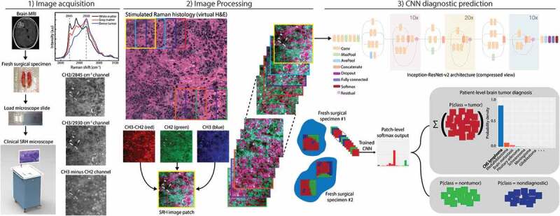
We recently developed and validated a bedside tissue-to-diagnosis pipeline using stimulated Raman histology (SRH), a label-free optical imaging method, and deep convolutional neural networks (CNN) in prospective clinical trial. Our CNN learned a hierarchy of interpretable histologic features found in the most common brain tumors and was able to accurately segment cancerous regions in SRH images.

 求助内容:
求助内容: 应助结果提醒方式:


