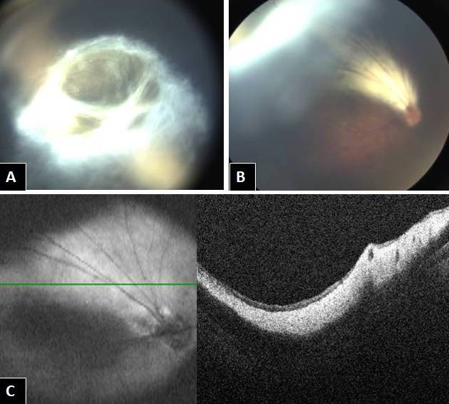
{"title":"网膜后纤维增生:下面是什么?","authors":"Parveen Sen, Dhaivat Shah","doi":"10.3205/oc000141","DOIUrl":null,"url":null,"abstract":"<p><p>A nine-month-old female baby with normal birth history presented with her mother complaining of a white spot in the baby's right eye, which the mother had noticed at five months of age. External photograph showed a retrolental fibroplastic membrane visible in the superior half of the dilated pupil. Retcam fundus photo revealed myelinated nerve fibers extending from the disc till the ora superiorly and forming a membranous fold. Intraoperative OCT confirmed thickened RNFL with compact retina. Thus, the retrolental fibroplasia turned out to be a masquerade for myelinated nerve fibers. Since it was not involving the visual axis with no coexisting traction, the mother was reassured regarding the benign nature of the condition.</p>","PeriodicalId":73178,"journal":{"name":"GMS ophthalmology cases","volume":"10 ","pages":"Doc14"},"PeriodicalIF":0.0000,"publicationDate":"2020-03-18","publicationTypes":"Journal Article","fieldsOfStudy":null,"isOpenAccess":false,"openAccessPdf":"https://www.ncbi.nlm.nih.gov/pmc/articles/PMC7114638/pdf/","citationCount":"1","resultStr":"{\"title\":\"Retrolental fibroplasias: What lies beneath?\",\"authors\":\"Parveen Sen, Dhaivat Shah\",\"doi\":\"10.3205/oc000141\",\"DOIUrl\":null,\"url\":null,\"abstract\":\"<p><p>A nine-month-old female baby with normal birth history presented with her mother complaining of a white spot in the baby's right eye, which the mother had noticed at five months of age. External photograph showed a retrolental fibroplastic membrane visible in the superior half of the dilated pupil. Retcam fundus photo revealed myelinated nerve fibers extending from the disc till the ora superiorly and forming a membranous fold. Intraoperative OCT confirmed thickened RNFL with compact retina. Thus, the retrolental fibroplasia turned out to be a masquerade for myelinated nerve fibers. Since it was not involving the visual axis with no coexisting traction, the mother was reassured regarding the benign nature of the condition.</p>\",\"PeriodicalId\":73178,\"journal\":{\"name\":\"GMS ophthalmology cases\",\"volume\":\"10 \",\"pages\":\"Doc14\"},\"PeriodicalIF\":0.0000,\"publicationDate\":\"2020-03-18\",\"publicationTypes\":\"Journal Article\",\"fieldsOfStudy\":null,\"isOpenAccess\":false,\"openAccessPdf\":\"https://www.ncbi.nlm.nih.gov/pmc/articles/PMC7114638/pdf/\",\"citationCount\":\"1\",\"resultStr\":null,\"platform\":\"Semanticscholar\",\"paperid\":null,\"PeriodicalName\":\"GMS ophthalmology cases\",\"FirstCategoryId\":\"1085\",\"ListUrlMain\":\"https://doi.org/10.3205/oc000141\",\"RegionNum\":0,\"RegionCategory\":null,\"ArticlePicture\":[],\"TitleCN\":null,\"AbstractTextCN\":null,\"PMCID\":null,\"EPubDate\":\"2020/1/1 0:00:00\",\"PubModel\":\"eCollection\",\"JCR\":\"\",\"JCRName\":\"\",\"Score\":null,\"Total\":0}","platform":"Semanticscholar","paperid":null,"PeriodicalName":"GMS ophthalmology cases","FirstCategoryId":"1085","ListUrlMain":"https://doi.org/10.3205/oc000141","RegionNum":0,"RegionCategory":null,"ArticlePicture":[],"TitleCN":null,"AbstractTextCN":null,"PMCID":null,"EPubDate":"2020/1/1 0:00:00","PubModel":"eCollection","JCR":"","JCRName":"","Score":null,"Total":0}
A nine-month-old female baby with normal birth history presented with her mother complaining of a white spot in the baby's right eye, which the mother had noticed at five months of age. External photograph showed a retrolental fibroplastic membrane visible in the superior half of the dilated pupil. Retcam fundus photo revealed myelinated nerve fibers extending from the disc till the ora superiorly and forming a membranous fold. Intraoperative OCT confirmed thickened RNFL with compact retina. Thus, the retrolental fibroplasia turned out to be a masquerade for myelinated nerve fibers. Since it was not involving the visual axis with no coexisting traction, the mother was reassured regarding the benign nature of the condition.

 求助内容:
求助内容: 应助结果提醒方式:


