Yeonju Choi, Yong-Il Kim, Seong-Sik Kim, Soo-Byung Park, Woo-Sung Son, Sung-Hun Kim
{"title":"下颌后移位对咽气道间隙的直接影响:初步研究。","authors":"Yeonju Choi, Yong-Il Kim, Seong-Sik Kim, Soo-Byung Park, Woo-Sung Son, Sung-Hun Kim","doi":"10.4041/kjod.2020.50.2.129","DOIUrl":null,"url":null,"abstract":"<p><strong>Objective: </strong>This study aimed to evaluate the immediate effects of mandibular posterior displacement on the pharyngeal airway space (PAS) by using cephalometric evaluations and to investigate how the surrounding structures are schematically involved.</p><p><strong>Methods: </strong>In this retrospective study, 38 subjects with functional Class III malocclusion and two lateral cephalograms were selected. The first lateral cephalogram was taken with the mandible in the habitual occlusal position, and the second in anterior edge-to-edge bite. Paired <i>t</i>-test was used to analyze changes in the PAS, hyoid bone, tongue, and soft palate, followed by mandibular posterior displacement. Pearson's correlation analysis was used to determine the relationship between the amount of mandibular posterior displacement and other variables.</p><p><strong>Results: </strong>A statistically significant decrease was observed in the PAS following mandibular posterior displacement. Along with mandibular posterior displacement, the tongue decreased in length (<i>p</i> < 0.001) and increased in height (<i>p</i> < 0.05), while the soft palate increased in length, decreased in thickness, and was posteriorly displaced (<i>p</i> < 0.001). The hyoid bone was also posteriorly displaced (<i>p</i> < 0.05). There was no correlation between the amount of mandibular posterior displacement and the measured variables.</p><p><strong>Conclusions: </strong>The PAS showed a statistically significant decrease following mandibular posterior displacement, which was a consequence of retraction of the surrounding structures. However, there were individual variances between the amount of mandibular posterior displacement and the measured variables.</p>","PeriodicalId":49934,"journal":{"name":"Korean Journal of Orthodontics","volume":"50 2","pages":"129-135"},"PeriodicalIF":1.9000,"publicationDate":"2020-03-01","publicationTypes":"Journal Article","fieldsOfStudy":null,"isOpenAccess":false,"openAccessPdf":"https://ftp.ncbi.nlm.nih.gov/pub/pmc/oa_pdf/5b/8c/kjod-50-129.PMC7093667.pdf","citationCount":"3","resultStr":"{\"title\":\"Immediate effects of mandibular posterior displacement on the pharyngeal airway space: A preliminary study.\",\"authors\":\"Yeonju Choi, Yong-Il Kim, Seong-Sik Kim, Soo-Byung Park, Woo-Sung Son, Sung-Hun Kim\",\"doi\":\"10.4041/kjod.2020.50.2.129\",\"DOIUrl\":null,\"url\":null,\"abstract\":\"<p><strong>Objective: </strong>This study aimed to evaluate the immediate effects of mandibular posterior displacement on the pharyngeal airway space (PAS) by using cephalometric evaluations and to investigate how the surrounding structures are schematically involved.</p><p><strong>Methods: </strong>In this retrospective study, 38 subjects with functional Class III malocclusion and two lateral cephalograms were selected. The first lateral cephalogram was taken with the mandible in the habitual occlusal position, and the second in anterior edge-to-edge bite. Paired <i>t</i>-test was used to analyze changes in the PAS, hyoid bone, tongue, and soft palate, followed by mandibular posterior displacement. Pearson's correlation analysis was used to determine the relationship between the amount of mandibular posterior displacement and other variables.</p><p><strong>Results: </strong>A statistically significant decrease was observed in the PAS following mandibular posterior displacement. Along with mandibular posterior displacement, the tongue decreased in length (<i>p</i> < 0.001) and increased in height (<i>p</i> < 0.05), while the soft palate increased in length, decreased in thickness, and was posteriorly displaced (<i>p</i> < 0.001). The hyoid bone was also posteriorly displaced (<i>p</i> < 0.05). There was no correlation between the amount of mandibular posterior displacement and the measured variables.</p><p><strong>Conclusions: </strong>The PAS showed a statistically significant decrease following mandibular posterior displacement, which was a consequence of retraction of the surrounding structures. However, there were individual variances between the amount of mandibular posterior displacement and the measured variables.</p>\",\"PeriodicalId\":49934,\"journal\":{\"name\":\"Korean Journal of Orthodontics\",\"volume\":\"50 2\",\"pages\":\"129-135\"},\"PeriodicalIF\":1.9000,\"publicationDate\":\"2020-03-01\",\"publicationTypes\":\"Journal Article\",\"fieldsOfStudy\":null,\"isOpenAccess\":false,\"openAccessPdf\":\"https://ftp.ncbi.nlm.nih.gov/pub/pmc/oa_pdf/5b/8c/kjod-50-129.PMC7093667.pdf\",\"citationCount\":\"3\",\"resultStr\":null,\"platform\":\"Semanticscholar\",\"paperid\":null,\"PeriodicalName\":\"Korean Journal of Orthodontics\",\"FirstCategoryId\":\"3\",\"ListUrlMain\":\"https://doi.org/10.4041/kjod.2020.50.2.129\",\"RegionNum\":3,\"RegionCategory\":\"医学\",\"ArticlePicture\":[],\"TitleCN\":null,\"AbstractTextCN\":null,\"PMCID\":null,\"EPubDate\":\"2020/3/24 0:00:00\",\"PubModel\":\"Epub\",\"JCR\":\"Q1\",\"JCRName\":\"Dentistry\",\"Score\":null,\"Total\":0}","platform":"Semanticscholar","paperid":null,"PeriodicalName":"Korean Journal of Orthodontics","FirstCategoryId":"3","ListUrlMain":"https://doi.org/10.4041/kjod.2020.50.2.129","RegionNum":3,"RegionCategory":"医学","ArticlePicture":[],"TitleCN":null,"AbstractTextCN":null,"PMCID":null,"EPubDate":"2020/3/24 0:00:00","PubModel":"Epub","JCR":"Q1","JCRName":"Dentistry","Score":null,"Total":0}
Immediate effects of mandibular posterior displacement on the pharyngeal airway space: A preliminary study.
Objective: This study aimed to evaluate the immediate effects of mandibular posterior displacement on the pharyngeal airway space (PAS) by using cephalometric evaluations and to investigate how the surrounding structures are schematically involved.
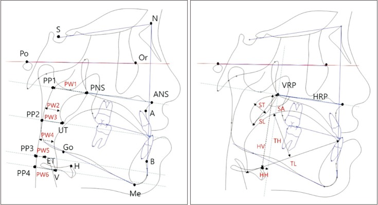
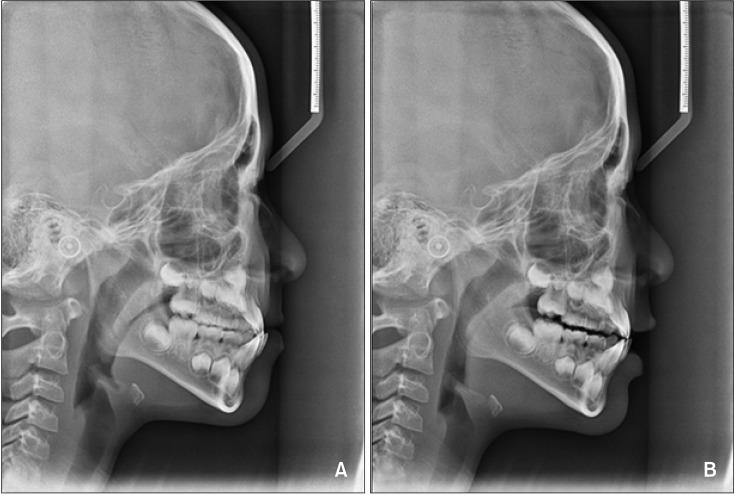
Methods: In this retrospective study, 38 subjects with functional Class III malocclusion and two lateral cephalograms were selected. The first lateral cephalogram was taken with the mandible in the habitual occlusal position, and the second in anterior edge-to-edge bite. Paired t-test was used to analyze changes in the PAS, hyoid bone, tongue, and soft palate, followed by mandibular posterior displacement. Pearson's correlation analysis was used to determine the relationship between the amount of mandibular posterior displacement and other variables.
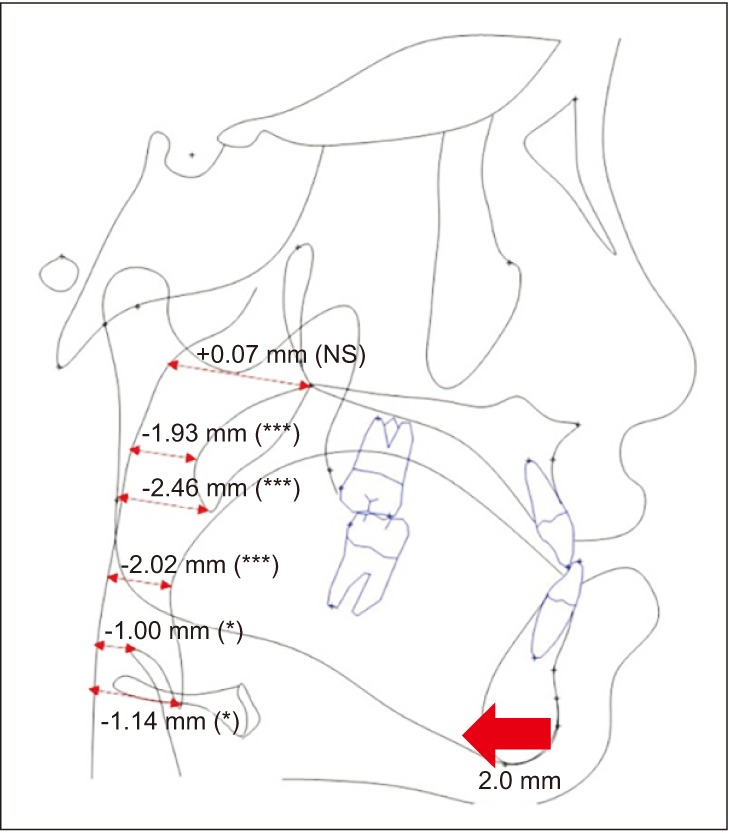
Results: A statistically significant decrease was observed in the PAS following mandibular posterior displacement. Along with mandibular posterior displacement, the tongue decreased in length (p < 0.001) and increased in height (p < 0.05), while the soft palate increased in length, decreased in thickness, and was posteriorly displaced (p < 0.001). The hyoid bone was also posteriorly displaced (p < 0.05). There was no correlation between the amount of mandibular posterior displacement and the measured variables.
Conclusions: The PAS showed a statistically significant decrease following mandibular posterior displacement, which was a consequence of retraction of the surrounding structures. However, there were individual variances between the amount of mandibular posterior displacement and the measured variables.
期刊介绍:
The Korean Journal of Orthodontics (KJO) is an international, open access, peer reviewed journal published in January, March, May, July, September, and November each year. It was first launched in 1970 and, as the official scientific publication of Korean Association of Orthodontists, KJO aims to publish high quality clinical and scientific original research papers in all areas related to orthodontics and dentofacial orthopedics. Specifically, its interest focuses on evidence-based investigations of contemporary diagnostic procedures and treatment techniques, expanding to significant clinical reports of diverse treatment approaches.
The scope of KJO covers all areas of orthodontics and dentofacial orthopedics including successful diagnostic procedures and treatment planning, growth and development of the face and its clinical implications, appliance designs, biomechanics, TMJ disorders and adult treatment. Specifically, its latest interest focuses on skeletal anchorage devices, orthodontic appliance and biomaterials, 3 dimensional imaging techniques utilized for dentofacial diagnosis and treatment planning, and orthognathic surgery to correct skeletal disharmony in association of orthodontic treatment.
 求助内容:
求助内容: 应助结果提醒方式:


