Yuan-Yuan Jiang, Lian Sun, Hua Wang, Chun-Yang Zhao, Wei-Bing Zhang
{"title":"三维锥束计算机断层扫描分析颞下颌关节对双块功能矫治器的反应。","authors":"Yuan-Yuan Jiang, Lian Sun, Hua Wang, Chun-Yang Zhao, Wei-Bing Zhang","doi":"10.4041/kjod.2020.50.2.86","DOIUrl":null,"url":null,"abstract":"<p><strong>Objective: </strong>To propose a three-dimensional (3D) method for evaluating temporomandibular joint (TMJ) changes during Twin-block treatment.</p><p><strong>Methods: </strong>Seventeen patients with Class II division 1 malocclusion treated using Twin-block and nine untreated patients with a similar malocclusion were included in this research. We collected their cone beam computed tomography (CBCT) data from before and 8 months after treatment. Segmentations were constructed using ITK-SNAP. Condylar volume and superficial area were measured using 3D Slicer. The 3D landmarks were identified on CBCT images by using Dolphin software to assess the condylar positional relationship. 3D models of the mandible and glenoid fossa of the patients were constructed and registered via voxel-based superimposition using 3D Slicer. Thereafter, skeletal changes could be visualized using 3DMeshMetric in any direction of the superimposition on a color-coded map. All the superimpositions were measured using the same scale on the distance color-coded map, in which red color represents overgrowth and blue color represents resorption.</p><p><strong>Results: </strong>Significant differences were observed in condylar volume, superficial area, and condylar position in both groups after 8 months. Compared with the control group (CG), the Twin-block group exhibited more obvious condyle-fossa modifications and joint positional changes. Moreover, on the color-coded map, more obvious condyle-fossa modifications could be observed in the posterior and superior directions in the Twin-block group than in the CG.</p><p><strong>Conclusions: </strong>We successfully established a 3D method for measuring and evaluating TMJ changes caused by Twin-block treatment. The treatment produced a larger condylar size and caused condylar positional changes.</p>","PeriodicalId":49934,"journal":{"name":"Korean Journal of Orthodontics","volume":"50 2","pages":"86-97"},"PeriodicalIF":1.9000,"publicationDate":"2020-03-01","publicationTypes":"Journal Article","fieldsOfStudy":null,"isOpenAccess":false,"openAccessPdf":"https://ftp.ncbi.nlm.nih.gov/pub/pmc/oa_pdf/83/a5/kjod-50-86.PMC7093662.pdf","citationCount":"12","resultStr":"{\"title\":\"Three-dimensional cone beam computed tomography analysis of temporomandibular joint response to the Twin-block functional appliance.\",\"authors\":\"Yuan-Yuan Jiang, Lian Sun, Hua Wang, Chun-Yang Zhao, Wei-Bing Zhang\",\"doi\":\"10.4041/kjod.2020.50.2.86\",\"DOIUrl\":null,\"url\":null,\"abstract\":\"<p><strong>Objective: </strong>To propose a three-dimensional (3D) method for evaluating temporomandibular joint (TMJ) changes during Twin-block treatment.</p><p><strong>Methods: </strong>Seventeen patients with Class II division 1 malocclusion treated using Twin-block and nine untreated patients with a similar malocclusion were included in this research. We collected their cone beam computed tomography (CBCT) data from before and 8 months after treatment. Segmentations were constructed using ITK-SNAP. Condylar volume and superficial area were measured using 3D Slicer. The 3D landmarks were identified on CBCT images by using Dolphin software to assess the condylar positional relationship. 3D models of the mandible and glenoid fossa of the patients were constructed and registered via voxel-based superimposition using 3D Slicer. Thereafter, skeletal changes could be visualized using 3DMeshMetric in any direction of the superimposition on a color-coded map. All the superimpositions were measured using the same scale on the distance color-coded map, in which red color represents overgrowth and blue color represents resorption.</p><p><strong>Results: </strong>Significant differences were observed in condylar volume, superficial area, and condylar position in both groups after 8 months. Compared with the control group (CG), the Twin-block group exhibited more obvious condyle-fossa modifications and joint positional changes. Moreover, on the color-coded map, more obvious condyle-fossa modifications could be observed in the posterior and superior directions in the Twin-block group than in the CG.</p><p><strong>Conclusions: </strong>We successfully established a 3D method for measuring and evaluating TMJ changes caused by Twin-block treatment. The treatment produced a larger condylar size and caused condylar positional changes.</p>\",\"PeriodicalId\":49934,\"journal\":{\"name\":\"Korean Journal of Orthodontics\",\"volume\":\"50 2\",\"pages\":\"86-97\"},\"PeriodicalIF\":1.9000,\"publicationDate\":\"2020-03-01\",\"publicationTypes\":\"Journal Article\",\"fieldsOfStudy\":null,\"isOpenAccess\":false,\"openAccessPdf\":\"https://ftp.ncbi.nlm.nih.gov/pub/pmc/oa_pdf/83/a5/kjod-50-86.PMC7093662.pdf\",\"citationCount\":\"12\",\"resultStr\":null,\"platform\":\"Semanticscholar\",\"paperid\":null,\"PeriodicalName\":\"Korean Journal of Orthodontics\",\"FirstCategoryId\":\"3\",\"ListUrlMain\":\"https://doi.org/10.4041/kjod.2020.50.2.86\",\"RegionNum\":3,\"RegionCategory\":\"医学\",\"ArticlePicture\":[],\"TitleCN\":null,\"AbstractTextCN\":null,\"PMCID\":null,\"EPubDate\":\"2020/3/24 0:00:00\",\"PubModel\":\"Epub\",\"JCR\":\"Q1\",\"JCRName\":\"Dentistry\",\"Score\":null,\"Total\":0}","platform":"Semanticscholar","paperid":null,"PeriodicalName":"Korean Journal of Orthodontics","FirstCategoryId":"3","ListUrlMain":"https://doi.org/10.4041/kjod.2020.50.2.86","RegionNum":3,"RegionCategory":"医学","ArticlePicture":[],"TitleCN":null,"AbstractTextCN":null,"PMCID":null,"EPubDate":"2020/3/24 0:00:00","PubModel":"Epub","JCR":"Q1","JCRName":"Dentistry","Score":null,"Total":0}
Three-dimensional cone beam computed tomography analysis of temporomandibular joint response to the Twin-block functional appliance.
Objective: To propose a three-dimensional (3D) method for evaluating temporomandibular joint (TMJ) changes during Twin-block treatment.
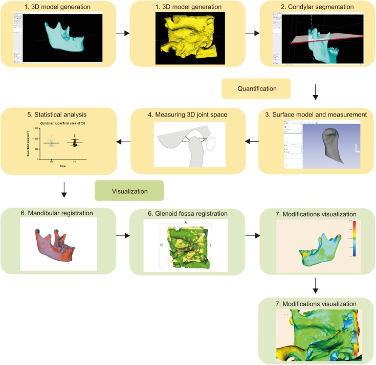
Methods: Seventeen patients with Class II division 1 malocclusion treated using Twin-block and nine untreated patients with a similar malocclusion were included in this research. We collected their cone beam computed tomography (CBCT) data from before and 8 months after treatment. Segmentations were constructed using ITK-SNAP. Condylar volume and superficial area were measured using 3D Slicer. The 3D landmarks were identified on CBCT images by using Dolphin software to assess the condylar positional relationship. 3D models of the mandible and glenoid fossa of the patients were constructed and registered via voxel-based superimposition using 3D Slicer. Thereafter, skeletal changes could be visualized using 3DMeshMetric in any direction of the superimposition on a color-coded map. All the superimpositions were measured using the same scale on the distance color-coded map, in which red color represents overgrowth and blue color represents resorption.
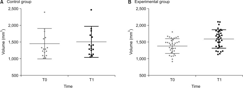
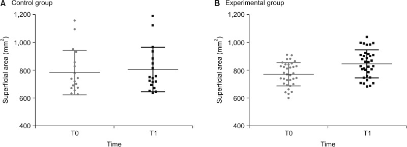
Results: Significant differences were observed in condylar volume, superficial area, and condylar position in both groups after 8 months. Compared with the control group (CG), the Twin-block group exhibited more obvious condyle-fossa modifications and joint positional changes. Moreover, on the color-coded map, more obvious condyle-fossa modifications could be observed in the posterior and superior directions in the Twin-block group than in the CG.
Conclusions: We successfully established a 3D method for measuring and evaluating TMJ changes caused by Twin-block treatment. The treatment produced a larger condylar size and caused condylar positional changes.
期刊介绍:
The Korean Journal of Orthodontics (KJO) is an international, open access, peer reviewed journal published in January, March, May, July, September, and November each year. It was first launched in 1970 and, as the official scientific publication of Korean Association of Orthodontists, KJO aims to publish high quality clinical and scientific original research papers in all areas related to orthodontics and dentofacial orthopedics. Specifically, its interest focuses on evidence-based investigations of contemporary diagnostic procedures and treatment techniques, expanding to significant clinical reports of diverse treatment approaches.
The scope of KJO covers all areas of orthodontics and dentofacial orthopedics including successful diagnostic procedures and treatment planning, growth and development of the face and its clinical implications, appliance designs, biomechanics, TMJ disorders and adult treatment. Specifically, its latest interest focuses on skeletal anchorage devices, orthodontic appliance and biomaterials, 3 dimensional imaging techniques utilized for dentofacial diagnosis and treatment planning, and orthognathic surgery to correct skeletal disharmony in association of orthodontic treatment.
 求助内容:
求助内容: 应助结果提醒方式:


