{"title":"微血管壁细胞自发Ca2+活性的同步性。","authors":"Retsu Mitsui, Hikaru Hashitani","doi":"10.1540/jsmr.56.1","DOIUrl":null,"url":null,"abstract":"<p><p>Spontaneous rhythmic constrictions known as vasomotion are developed in several microvascular beds in vivo. Vasomotion in arterioles is considered to facilitate blood flow, while venular vasomotion would facilitate tissue metabolite drainage. Mechanisms underlying vasomotion periodically generate synchronous Ca<sup>2+</sup> transients in vascular smooth muscle cells (VSMCs). In visceral organs, mural cells (pericytes and VSMCs) in arterioles, capillaries and venules exhibit synchronous spontaneous Ca<sup>2+</sup> transients. Since sympathetic regulation is rather limited in the intra-organ microvessels, spontaneous activity of mural cells may play an essential role in maintaining tissue perfusion. Synchronous spontaneous Ca<sup>2+</sup> transients in precapillary arterioles (PCAs)/capillaries appear to propagate to upstream arterioles to drive their vasomotion, while venules develop their own synchronous Ca<sup>2+</sup> transients and associated vasomotion. Spontaneous Ca<sup>2+</sup> transients of mural cells primarily arise from IP<sub>3</sub> and/or ryanodine receptor-mediated Ca<sup>2+</sup> release from sarcoendoplasmic reticulum (SR/ER) Ca<sup>2+</sup> stores. The resultant opening of Ca<sup>2+</sup>-activated Cl<sup>-</sup> channels (CaCCs) causes a membrane depolarisation that triggers Ca<sup>2+</sup> influx via T-type and/or L-type voltage-dependent Ca<sup>2+</sup> channels (VDCCs). Mural cells are electrically coupled with each other via gap junctions, and thus allow the sequential spread of CaCC or VDCC-dependent depolarisations to develop the synchrony of Ca<sup>2+</sup> transients within their network. Importantly, the synchrony of spontaneous Ca<sup>2+</sup> transients also requires a certain range of the resting membrane potential that is maintained by the opening of K<sub>v</sub>7 voltage-dependent K<sup>+</sup> (K<sub>v</sub>7) and inward rectifier K<sup>+</sup> (K<sub>ir</sub>) channels. Thus, a depolarised membrane would evoke asynchronous, 'premature' spontaneous Ca<sup>2+</sup> transients, while a hyperpolarised membrane prevents any spontaneous activity.</p>","PeriodicalId":39619,"journal":{"name":"Journal of Smooth Muscle Research","volume":"56 0","pages":"1-18"},"PeriodicalIF":0.0000,"publicationDate":"2020-01-01","publicationTypes":"Journal Article","fieldsOfStudy":null,"isOpenAccess":false,"openAccessPdf":"https://ftp.ncbi.nlm.nih.gov/pub/pmc/oa_pdf/2e/d2/jsmr-56-001.PMC7132055.pdf","citationCount":"5","resultStr":"{\"title\":\"Synchrony of spontaneous Ca<sup>2+</sup> activity in microvascular mural cells.\",\"authors\":\"Retsu Mitsui, Hikaru Hashitani\",\"doi\":\"10.1540/jsmr.56.1\",\"DOIUrl\":null,\"url\":null,\"abstract\":\"<p><p>Spontaneous rhythmic constrictions known as vasomotion are developed in several microvascular beds in vivo. Vasomotion in arterioles is considered to facilitate blood flow, while venular vasomotion would facilitate tissue metabolite drainage. Mechanisms underlying vasomotion periodically generate synchronous Ca<sup>2+</sup> transients in vascular smooth muscle cells (VSMCs). In visceral organs, mural cells (pericytes and VSMCs) in arterioles, capillaries and venules exhibit synchronous spontaneous Ca<sup>2+</sup> transients. Since sympathetic regulation is rather limited in the intra-organ microvessels, spontaneous activity of mural cells may play an essential role in maintaining tissue perfusion. Synchronous spontaneous Ca<sup>2+</sup> transients in precapillary arterioles (PCAs)/capillaries appear to propagate to upstream arterioles to drive their vasomotion, while venules develop their own synchronous Ca<sup>2+</sup> transients and associated vasomotion. Spontaneous Ca<sup>2+</sup> transients of mural cells primarily arise from IP<sub>3</sub> and/or ryanodine receptor-mediated Ca<sup>2+</sup> release from sarcoendoplasmic reticulum (SR/ER) Ca<sup>2+</sup> stores. The resultant opening of Ca<sup>2+</sup>-activated Cl<sup>-</sup> channels (CaCCs) causes a membrane depolarisation that triggers Ca<sup>2+</sup> influx via T-type and/or L-type voltage-dependent Ca<sup>2+</sup> channels (VDCCs). Mural cells are electrically coupled with each other via gap junctions, and thus allow the sequential spread of CaCC or VDCC-dependent depolarisations to develop the synchrony of Ca<sup>2+</sup> transients within their network. Importantly, the synchrony of spontaneous Ca<sup>2+</sup> transients also requires a certain range of the resting membrane potential that is maintained by the opening of K<sub>v</sub>7 voltage-dependent K<sup>+</sup> (K<sub>v</sub>7) and inward rectifier K<sup>+</sup> (K<sub>ir</sub>) channels. Thus, a depolarised membrane would evoke asynchronous, 'premature' spontaneous Ca<sup>2+</sup> transients, while a hyperpolarised membrane prevents any spontaneous activity.</p>\",\"PeriodicalId\":39619,\"journal\":{\"name\":\"Journal of Smooth Muscle Research\",\"volume\":\"56 0\",\"pages\":\"1-18\"},\"PeriodicalIF\":0.0000,\"publicationDate\":\"2020-01-01\",\"publicationTypes\":\"Journal Article\",\"fieldsOfStudy\":null,\"isOpenAccess\":false,\"openAccessPdf\":\"https://ftp.ncbi.nlm.nih.gov/pub/pmc/oa_pdf/2e/d2/jsmr-56-001.PMC7132055.pdf\",\"citationCount\":\"5\",\"resultStr\":null,\"platform\":\"Semanticscholar\",\"paperid\":null,\"PeriodicalName\":\"Journal of Smooth Muscle Research\",\"FirstCategoryId\":\"1085\",\"ListUrlMain\":\"https://doi.org/10.1540/jsmr.56.1\",\"RegionNum\":0,\"RegionCategory\":null,\"ArticlePicture\":[],\"TitleCN\":null,\"AbstractTextCN\":null,\"PMCID\":null,\"EPubDate\":\"\",\"PubModel\":\"\",\"JCR\":\"Q3\",\"JCRName\":\"Medicine\",\"Score\":null,\"Total\":0}","platform":"Semanticscholar","paperid":null,"PeriodicalName":"Journal of Smooth Muscle Research","FirstCategoryId":"1085","ListUrlMain":"https://doi.org/10.1540/jsmr.56.1","RegionNum":0,"RegionCategory":null,"ArticlePicture":[],"TitleCN":null,"AbstractTextCN":null,"PMCID":null,"EPubDate":"","PubModel":"","JCR":"Q3","JCRName":"Medicine","Score":null,"Total":0}
Synchrony of spontaneous Ca2+ activity in microvascular mural cells.
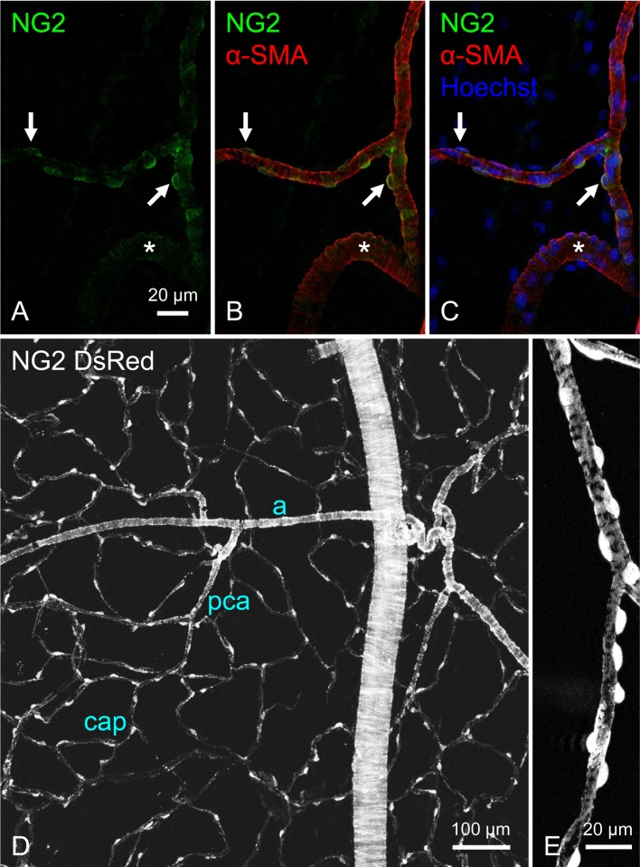
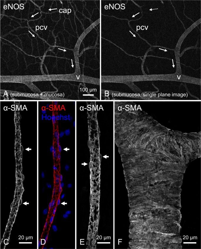
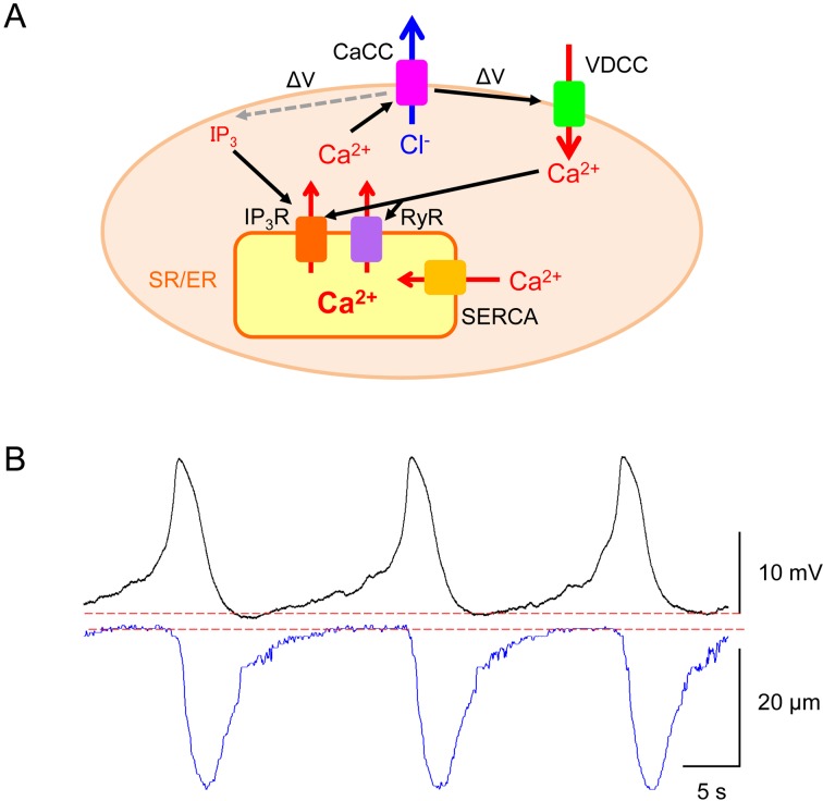
Spontaneous rhythmic constrictions known as vasomotion are developed in several microvascular beds in vivo. Vasomotion in arterioles is considered to facilitate blood flow, while venular vasomotion would facilitate tissue metabolite drainage. Mechanisms underlying vasomotion periodically generate synchronous Ca2+ transients in vascular smooth muscle cells (VSMCs). In visceral organs, mural cells (pericytes and VSMCs) in arterioles, capillaries and venules exhibit synchronous spontaneous Ca2+ transients. Since sympathetic regulation is rather limited in the intra-organ microvessels, spontaneous activity of mural cells may play an essential role in maintaining tissue perfusion. Synchronous spontaneous Ca2+ transients in precapillary arterioles (PCAs)/capillaries appear to propagate to upstream arterioles to drive their vasomotion, while venules develop their own synchronous Ca2+ transients and associated vasomotion. Spontaneous Ca2+ transients of mural cells primarily arise from IP3 and/or ryanodine receptor-mediated Ca2+ release from sarcoendoplasmic reticulum (SR/ER) Ca2+ stores. The resultant opening of Ca2+-activated Cl- channels (CaCCs) causes a membrane depolarisation that triggers Ca2+ influx via T-type and/or L-type voltage-dependent Ca2+ channels (VDCCs). Mural cells are electrically coupled with each other via gap junctions, and thus allow the sequential spread of CaCC or VDCC-dependent depolarisations to develop the synchrony of Ca2+ transients within their network. Importantly, the synchrony of spontaneous Ca2+ transients also requires a certain range of the resting membrane potential that is maintained by the opening of Kv7 voltage-dependent K+ (Kv7) and inward rectifier K+ (Kir) channels. Thus, a depolarised membrane would evoke asynchronous, 'premature' spontaneous Ca2+ transients, while a hyperpolarised membrane prevents any spontaneous activity.

 求助内容:
求助内容: 应助结果提醒方式:


