Amer F. Saleh, Elisa Lázaro-Ibáñez, Malin A.-M. Forsgard, Olga Shatnyeva, Xabier Osteikoetxea, Fredrik Karlsson, Nikki Heath, Madeleine Ingelsten, Jonathan Rose, Jayne Harris, Maelle Mairesse, Stephanie M. Bates, Maryam Clausen, Damla Etal, Emilyanne Leonard, Mick D. Fellows, Niek Dekker and Nicholas Edmunds
{"title":"细胞外囊泡诱导最小肝毒性和免疫原性†","authors":"Amer F. Saleh, Elisa Lázaro-Ibáñez, Malin A.-M. Forsgard, Olga Shatnyeva, Xabier Osteikoetxea, Fredrik Karlsson, Nikki Heath, Madeleine Ingelsten, Jonathan Rose, Jayne Harris, Maelle Mairesse, Stephanie M. Bates, Maryam Clausen, Damla Etal, Emilyanne Leonard, Mick D. Fellows, Niek Dekker and Nicholas Edmunds","doi":"10.1039/C8NR08720B","DOIUrl":null,"url":null,"abstract":"<p >Extracellular vesicles (EVs) mediate cellular communication through the transfer of active biomolecules, raising interest in using them as biological delivery vehicles for therapeutic drugs. For drug delivery applications, it is important to understand the intrinsic safety and toxicity liabilities of EVs. Nanoparticles, including EVs, typically demonstrate significant accumulation in the liver after systemic administration <em>in vivo</em>. We confirmed uptake of EVs derived from Expi293F cells into HepG2 cells and did not detect any signs of hepatotoxicity measured by cell viability, functional secretion of albumin, plasma membrane integrity, and mitochondrial and lysosomal activity even at high exposures of up to 5 × 10<small><sup>10</sup></small> EVs per mL. Whole genome transcriptome analysis was used to measure potential effects on the gene expression in the recipient HepG2 cells at 24 h following exposure to EVs. Only 0.6% of all genes were found to be differentially expressed displaying less than 2-fold expression change, with genes related to inflammation or toxicity being unaffected. EVs did not trigger any proinflammatory cytokine response in HepG2 cells. However, minor changes were noted in human blood for interleukin (IL)-8, IL-6, and monocyte chemotactic protein 1 (MCP-1). Administration of 5 × 10<small><sup>10</sup></small> Expi293F-derived EVs to BALB/c mice did not result in any histopathological changes or increases of liver transaminases or cytokine levels, apart from a modest increase in keratinocyte chemoattractant (KC). The absence of any significant toxicity associated with EVs <em>in vitro</em> and <em>in vivo</em> supports the prospective use of EVs for therapeutic applications and for drug delivery.</p>","PeriodicalId":92,"journal":{"name":"Nanoscale","volume":" 14","pages":" 6990-7001"},"PeriodicalIF":5.8000,"publicationDate":"2019-03-07","publicationTypes":"Journal Article","fieldsOfStudy":null,"isOpenAccess":false,"openAccessPdf":"https://sci-hub-pdf.com/10.1039/C8NR08720B","citationCount":"95","resultStr":"{\"title\":\"Extracellular vesicles induce minimal hepatotoxicity and immunogenicity†\",\"authors\":\"Amer F. Saleh, Elisa Lázaro-Ibáñez, Malin A.-M. Forsgard, Olga Shatnyeva, Xabier Osteikoetxea, Fredrik Karlsson, Nikki Heath, Madeleine Ingelsten, Jonathan Rose, Jayne Harris, Maelle Mairesse, Stephanie M. Bates, Maryam Clausen, Damla Etal, Emilyanne Leonard, Mick D. Fellows, Niek Dekker and Nicholas Edmunds\",\"doi\":\"10.1039/C8NR08720B\",\"DOIUrl\":null,\"url\":null,\"abstract\":\"<p >Extracellular vesicles (EVs) mediate cellular communication through the transfer of active biomolecules, raising interest in using them as biological delivery vehicles for therapeutic drugs. For drug delivery applications, it is important to understand the intrinsic safety and toxicity liabilities of EVs. Nanoparticles, including EVs, typically demonstrate significant accumulation in the liver after systemic administration <em>in vivo</em>. We confirmed uptake of EVs derived from Expi293F cells into HepG2 cells and did not detect any signs of hepatotoxicity measured by cell viability, functional secretion of albumin, plasma membrane integrity, and mitochondrial and lysosomal activity even at high exposures of up to 5 × 10<small><sup>10</sup></small> EVs per mL. Whole genome transcriptome analysis was used to measure potential effects on the gene expression in the recipient HepG2 cells at 24 h following exposure to EVs. Only 0.6% of all genes were found to be differentially expressed displaying less than 2-fold expression change, with genes related to inflammation or toxicity being unaffected. EVs did not trigger any proinflammatory cytokine response in HepG2 cells. However, minor changes were noted in human blood for interleukin (IL)-8, IL-6, and monocyte chemotactic protein 1 (MCP-1). Administration of 5 × 10<small><sup>10</sup></small> Expi293F-derived EVs to BALB/c mice did not result in any histopathological changes or increases of liver transaminases or cytokine levels, apart from a modest increase in keratinocyte chemoattractant (KC). The absence of any significant toxicity associated with EVs <em>in vitro</em> and <em>in vivo</em> supports the prospective use of EVs for therapeutic applications and for drug delivery.</p>\",\"PeriodicalId\":92,\"journal\":{\"name\":\"Nanoscale\",\"volume\":\" 14\",\"pages\":\" 6990-7001\"},\"PeriodicalIF\":5.8000,\"publicationDate\":\"2019-03-07\",\"publicationTypes\":\"Journal Article\",\"fieldsOfStudy\":null,\"isOpenAccess\":false,\"openAccessPdf\":\"https://sci-hub-pdf.com/10.1039/C8NR08720B\",\"citationCount\":\"95\",\"resultStr\":null,\"platform\":\"Semanticscholar\",\"paperid\":null,\"PeriodicalName\":\"Nanoscale\",\"FirstCategoryId\":\"88\",\"ListUrlMain\":\"https://pubs.rsc.org/en/content/articlelanding/2019/nr/c8nr08720b\",\"RegionNum\":3,\"RegionCategory\":\"材料科学\",\"ArticlePicture\":[],\"TitleCN\":null,\"AbstractTextCN\":null,\"PMCID\":null,\"EPubDate\":\"\",\"PubModel\":\"\",\"JCR\":\"Q1\",\"JCRName\":\"CHEMISTRY, MULTIDISCIPLINARY\",\"Score\":null,\"Total\":0}","platform":"Semanticscholar","paperid":null,"PeriodicalName":"Nanoscale","FirstCategoryId":"88","ListUrlMain":"https://pubs.rsc.org/en/content/articlelanding/2019/nr/c8nr08720b","RegionNum":3,"RegionCategory":"材料科学","ArticlePicture":[],"TitleCN":null,"AbstractTextCN":null,"PMCID":null,"EPubDate":"","PubModel":"","JCR":"Q1","JCRName":"CHEMISTRY, MULTIDISCIPLINARY","Score":null,"Total":0}
引用次数: 95
摘要
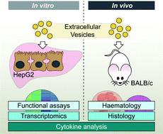
细胞外囊泡(EVs)通过活性生物分子的转移介导细胞通讯,这引起了人们对使用它们作为治疗药物的生物递送载体的兴趣。对于药物递送应用,了解电动汽车的内在安全性和毒性是很重要的。在体内全身给药后,纳米颗粒,包括ev,通常在肝脏中表现出显著的积累。我们证实了来自Expi293F细胞的ev被摄取到HepG2细胞中,并且即使在高达5 × 10¹⁰ ev / mL的高暴露下,通过细胞活力、白蛋白功能分泌、质膜完整性、线粒体和溶酶体活性测量,也没有检测到任何肝毒性的迹象。全基因组转录组分析用于测量ev暴露后24小时对受体HepG2细胞基因表达的潜在影响。只有0.6%的基因被发现存在差异表达,表达变化小于2倍,与炎症或毒性相关的基因未受影响。在HepG2细胞中,ev没有触发任何促炎细胞因子反应。然而,人血液中白细胞介素(IL)-8、IL-6和单核细胞趋化蛋白1 (MCP-1)的变化较小。5 × 10¹⁰ expi293f衍生ev给BALB/c小鼠,除了角质细胞化学引诱剂(KC)的适度增加外,没有导致任何组织病理学改变或肝转氨酶或细胞因子水平的增加。体外和体内均没有与ev相关的任何显著毒性,这支持了ev用于治疗和药物输送的前景。
Extracellular vesicles induce minimal hepatotoxicity and immunogenicity†
Extracellular vesicles (EVs) mediate cellular communication through the transfer of active biomolecules, raising interest in using them as biological delivery vehicles for therapeutic drugs. For drug delivery applications, it is important to understand the intrinsic safety and toxicity liabilities of EVs. Nanoparticles, including EVs, typically demonstrate significant accumulation in the liver after systemic administration in vivo. We confirmed uptake of EVs derived from Expi293F cells into HepG2 cells and did not detect any signs of hepatotoxicity measured by cell viability, functional secretion of albumin, plasma membrane integrity, and mitochondrial and lysosomal activity even at high exposures of up to 5 × 10¹⁰ EVs per mL. Whole genome transcriptome analysis was used to measure potential effects on the gene expression in the recipient HepG2 cells at 24 h following exposure to EVs. Only 0.6% of all genes were found to be differentially expressed displaying less than 2-fold expression change, with genes related to inflammation or toxicity being unaffected. EVs did not trigger any proinflammatory cytokine response in HepG2 cells. However, minor changes were noted in human blood for interleukin (IL)-8, IL-6, and monocyte chemotactic protein 1 (MCP-1). Administration of 5 × 10¹⁰ Expi293F-derived EVs to BALB/c mice did not result in any histopathological changes or increases of liver transaminases or cytokine levels, apart from a modest increase in keratinocyte chemoattractant (KC). The absence of any significant toxicity associated with EVs in vitro and in vivo supports the prospective use of EVs for therapeutic applications and for drug delivery.
期刊介绍:
Nanoscale is a high-impact international journal, publishing high-quality research across nanoscience and nanotechnology. Nanoscale publishes a full mix of research articles on experimental and theoretical work, including reviews, communications, and full papers.Highly interdisciplinary, this journal appeals to scientists, researchers and professionals interested in nanoscience and nanotechnology, quantum materials and quantum technology, including the areas of physics, chemistry, biology, medicine, materials, energy/environment, information technology, detection science, healthcare and drug discovery, and electronics.
 求助内容:
求助内容: 应助结果提醒方式:


