Chrysafis Andreou, Konstantinos Plakas, Naxhije Berisha, Mathieu Gigoux, Lauren E. Rosch, Rustin Mirsafavi, Anton Oseledchyk, Suchetan Pal, Dmitriy Zamarin, Taha Merghoub, Michael R. Detty and Moritz F. Kircher
{"title":"表面增强共振拉曼散射纳米探针的多路分子成像通过多通道图像分割显示小鼠免疫治疗应答","authors":"Chrysafis Andreou, Konstantinos Plakas, Naxhije Berisha, Mathieu Gigoux, Lauren E. Rosch, Rustin Mirsafavi, Anton Oseledchyk, Suchetan Pal, Dmitriy Zamarin, Taha Merghoub, Michael R. Detty and Moritz F. Kircher","doi":"10.1039/D2NH00331G","DOIUrl":null,"url":null,"abstract":"<p >Visualizing the presence and distribution of multiple specific molecular markers within a tumor can reveal the composition of its microenvironment, inform diagnosis, stratify patients, and guide treatment. Raman imaging with multiple molecularly-targeted surface enhanced Raman scattering (SERS) nanoprobes could help investigate emerging cancer treatments preclinically or enable personalized treatment assessment. Here, we report a comprehensive strategy for multiplexed imaging using SERS nanoprobes and machine learning (ML) to monitor the early effects of immune checkpoint blockade (ICB) in tumor-bearing mice. We used antibody-functionalized SERS nanoprobes to visualize 7 + 1 immunotherapy-related targets simultaneously. The multiplexed images were spectrally resolved and then spatially segmented into superpixels based on the unmixed signals. The superpixels were used to train ML models, leading to the successful classification of mice into treated and untreated groups, and identifying tumor regions with variable responses to treatment. This method may help predict treatment efficacy in tumors and identify areas of tumor variability and therapy resistance.</p>","PeriodicalId":93,"journal":{"name":"Nanoscale Horizons","volume":" 12","pages":" 1540-1552"},"PeriodicalIF":6.6000,"publicationDate":"2022-10-20","publicationTypes":"Journal Article","fieldsOfStudy":null,"isOpenAccess":false,"openAccessPdf":"https://pubs.rsc.org/en/content/articlepdf/2022/nh/d2nh00331g?page=search","citationCount":"0","resultStr":"{\"title\":\"Multiplexed molecular imaging with surface enhanced resonance Raman scattering nanoprobes reveals immunotherapy response in mice via multichannel image segmentation†\",\"authors\":\"Chrysafis Andreou, Konstantinos Plakas, Naxhije Berisha, Mathieu Gigoux, Lauren E. Rosch, Rustin Mirsafavi, Anton Oseledchyk, Suchetan Pal, Dmitriy Zamarin, Taha Merghoub, Michael R. Detty and Moritz F. Kircher\",\"doi\":\"10.1039/D2NH00331G\",\"DOIUrl\":null,\"url\":null,\"abstract\":\"<p >Visualizing the presence and distribution of multiple specific molecular markers within a tumor can reveal the composition of its microenvironment, inform diagnosis, stratify patients, and guide treatment. Raman imaging with multiple molecularly-targeted surface enhanced Raman scattering (SERS) nanoprobes could help investigate emerging cancer treatments preclinically or enable personalized treatment assessment. Here, we report a comprehensive strategy for multiplexed imaging using SERS nanoprobes and machine learning (ML) to monitor the early effects of immune checkpoint blockade (ICB) in tumor-bearing mice. We used antibody-functionalized SERS nanoprobes to visualize 7 + 1 immunotherapy-related targets simultaneously. The multiplexed images were spectrally resolved and then spatially segmented into superpixels based on the unmixed signals. The superpixels were used to train ML models, leading to the successful classification of mice into treated and untreated groups, and identifying tumor regions with variable responses to treatment. This method may help predict treatment efficacy in tumors and identify areas of tumor variability and therapy resistance.</p>\",\"PeriodicalId\":93,\"journal\":{\"name\":\"Nanoscale Horizons\",\"volume\":\" 12\",\"pages\":\" 1540-1552\"},\"PeriodicalIF\":6.6000,\"publicationDate\":\"2022-10-20\",\"publicationTypes\":\"Journal Article\",\"fieldsOfStudy\":null,\"isOpenAccess\":false,\"openAccessPdf\":\"https://pubs.rsc.org/en/content/articlepdf/2022/nh/d2nh00331g?page=search\",\"citationCount\":\"0\",\"resultStr\":null,\"platform\":\"Semanticscholar\",\"paperid\":null,\"PeriodicalName\":\"Nanoscale Horizons\",\"FirstCategoryId\":\"88\",\"ListUrlMain\":\"https://pubs.rsc.org/en/content/articlelanding/2022/nh/d2nh00331g\",\"RegionNum\":2,\"RegionCategory\":\"材料科学\",\"ArticlePicture\":[],\"TitleCN\":null,\"AbstractTextCN\":null,\"PMCID\":null,\"EPubDate\":\"\",\"PubModel\":\"\",\"JCR\":\"Q1\",\"JCRName\":\"CHEMISTRY, PHYSICAL\",\"Score\":null,\"Total\":0}","platform":"Semanticscholar","paperid":null,"PeriodicalName":"Nanoscale Horizons","FirstCategoryId":"88","ListUrlMain":"https://pubs.rsc.org/en/content/articlelanding/2022/nh/d2nh00331g","RegionNum":2,"RegionCategory":"材料科学","ArticlePicture":[],"TitleCN":null,"AbstractTextCN":null,"PMCID":null,"EPubDate":"","PubModel":"","JCR":"Q1","JCRName":"CHEMISTRY, PHYSICAL","Score":null,"Total":0}
Multiplexed molecular imaging with surface enhanced resonance Raman scattering nanoprobes reveals immunotherapy response in mice via multichannel image segmentation†
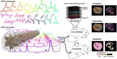
Visualizing the presence and distribution of multiple specific molecular markers within a tumor can reveal the composition of its microenvironment, inform diagnosis, stratify patients, and guide treatment. Raman imaging with multiple molecularly-targeted surface enhanced Raman scattering (SERS) nanoprobes could help investigate emerging cancer treatments preclinically or enable personalized treatment assessment. Here, we report a comprehensive strategy for multiplexed imaging using SERS nanoprobes and machine learning (ML) to monitor the early effects of immune checkpoint blockade (ICB) in tumor-bearing mice. We used antibody-functionalized SERS nanoprobes to visualize 7 + 1 immunotherapy-related targets simultaneously. The multiplexed images were spectrally resolved and then spatially segmented into superpixels based on the unmixed signals. The superpixels were used to train ML models, leading to the successful classification of mice into treated and untreated groups, and identifying tumor regions with variable responses to treatment. This method may help predict treatment efficacy in tumors and identify areas of tumor variability and therapy resistance.
期刊介绍:
Nanoscale Horizons stands out as a premier journal for publishing exceptionally high-quality and innovative nanoscience and nanotechnology. The emphasis lies on original research that introduces a new concept or a novel perspective (a conceptual advance), prioritizing this over reporting technological improvements. Nevertheless, outstanding articles showcasing truly groundbreaking developments, including record-breaking performance, may also find a place in the journal. Published work must be of substantial general interest to our broad and diverse readership across the nanoscience and nanotechnology community.
 求助内容:
求助内容: 应助结果提醒方式:


