{"title":"三维锥束ct头部重定向中矢状面比较。","authors":"Eon-Hwa Lee, Hyung-Seog Yu, Kee-Joon Lee, Sang-Sun Han, Hwi-Dong Jung, Chung-Ju Hwang","doi":"10.4041/kjod.2020.50.1.3","DOIUrl":null,"url":null,"abstract":"<p><strong>Objective: </strong>This study compared three prominent midsagittal planes (MSPs) to identify the MSP that best approximates the true symmetrical MSP.</p><p><strong>Methods: </strong>Forty-three patients (mean age, 23.0 ± 8.20 years) were grouped as follows: group 1 consisted of 10 patients with skeletal Class I and a menton (Me) deviation of < 2 mm; group 2, 11 patients with skeletal Class III and a Me deviation < 2 mm; group 3, nine patients with skeletal Class III and a Me deviation of 2 to less than 4 mm; and group 4, 13 patients with skeletal Class III and an Me deviation ≥ 4 mm. The candidate MSPs were established by three-dimensional (3D) cone beam computed tomography (CBCT) reorientation methods (RMs): (1) the MSP perpendicular to the Frankfort horizontal (FH) plane while passing through the crista galli and basion; (2) the MSP including the nasion, incisive foramen, and basion; (3) the MSP including the nasion, anterior nasal spine, and posterior nasal spine. The mean absolute distances (MADs) to the MSPs were calculated from the coordinates of 1,548 points on 129 CBCT images. The differences in the values of the 3D coordinates among RMs were compared.</p><p><strong>Results: </strong>The MADs of the three RMs showed significant differences (<i>p</i> < 0.05). Most of the differences in values of the coordinates were not significant among RMs.</p><p><strong>Conclusions: </strong>Although the differences in distance among the three MSPs were minor, the MSP perpendicular to the FH plane while passing through the crista galli and basion best approximated the true symmetrical MSP.</p>","PeriodicalId":49934,"journal":{"name":"Korean Journal of Orthodontics","volume":"50 1","pages":"3-12"},"PeriodicalIF":1.9000,"publicationDate":"2020-01-01","publicationTypes":"Journal Article","fieldsOfStudy":null,"isOpenAccess":false,"openAccessPdf":"https://ftp.ncbi.nlm.nih.gov/pub/pmc/oa_pdf/a4/05/kjod-50-3.PMC6995832.pdf","citationCount":"5","resultStr":"{\"title\":\"Comparison of three midsagittal planes for three-dimensional cone beam computed tomography head reorientation.\",\"authors\":\"Eon-Hwa Lee, Hyung-Seog Yu, Kee-Joon Lee, Sang-Sun Han, Hwi-Dong Jung, Chung-Ju Hwang\",\"doi\":\"10.4041/kjod.2020.50.1.3\",\"DOIUrl\":null,\"url\":null,\"abstract\":\"<p><strong>Objective: </strong>This study compared three prominent midsagittal planes (MSPs) to identify the MSP that best approximates the true symmetrical MSP.</p><p><strong>Methods: </strong>Forty-three patients (mean age, 23.0 ± 8.20 years) were grouped as follows: group 1 consisted of 10 patients with skeletal Class I and a menton (Me) deviation of < 2 mm; group 2, 11 patients with skeletal Class III and a Me deviation < 2 mm; group 3, nine patients with skeletal Class III and a Me deviation of 2 to less than 4 mm; and group 4, 13 patients with skeletal Class III and an Me deviation ≥ 4 mm. The candidate MSPs were established by three-dimensional (3D) cone beam computed tomography (CBCT) reorientation methods (RMs): (1) the MSP perpendicular to the Frankfort horizontal (FH) plane while passing through the crista galli and basion; (2) the MSP including the nasion, incisive foramen, and basion; (3) the MSP including the nasion, anterior nasal spine, and posterior nasal spine. The mean absolute distances (MADs) to the MSPs were calculated from the coordinates of 1,548 points on 129 CBCT images. The differences in the values of the 3D coordinates among RMs were compared.</p><p><strong>Results: </strong>The MADs of the three RMs showed significant differences (<i>p</i> < 0.05). Most of the differences in values of the coordinates were not significant among RMs.</p><p><strong>Conclusions: </strong>Although the differences in distance among the three MSPs were minor, the MSP perpendicular to the FH plane while passing through the crista galli and basion best approximated the true symmetrical MSP.</p>\",\"PeriodicalId\":49934,\"journal\":{\"name\":\"Korean Journal of Orthodontics\",\"volume\":\"50 1\",\"pages\":\"3-12\"},\"PeriodicalIF\":1.9000,\"publicationDate\":\"2020-01-01\",\"publicationTypes\":\"Journal Article\",\"fieldsOfStudy\":null,\"isOpenAccess\":false,\"openAccessPdf\":\"https://ftp.ncbi.nlm.nih.gov/pub/pmc/oa_pdf/a4/05/kjod-50-3.PMC6995832.pdf\",\"citationCount\":\"5\",\"resultStr\":null,\"platform\":\"Semanticscholar\",\"paperid\":null,\"PeriodicalName\":\"Korean Journal of Orthodontics\",\"FirstCategoryId\":\"3\",\"ListUrlMain\":\"https://doi.org/10.4041/kjod.2020.50.1.3\",\"RegionNum\":3,\"RegionCategory\":\"医学\",\"ArticlePicture\":[],\"TitleCN\":null,\"AbstractTextCN\":null,\"PMCID\":null,\"EPubDate\":\"2020/1/22 0:00:00\",\"PubModel\":\"Epub\",\"JCR\":\"Q1\",\"JCRName\":\"Dentistry\",\"Score\":null,\"Total\":0}","platform":"Semanticscholar","paperid":null,"PeriodicalName":"Korean Journal of Orthodontics","FirstCategoryId":"3","ListUrlMain":"https://doi.org/10.4041/kjod.2020.50.1.3","RegionNum":3,"RegionCategory":"医学","ArticlePicture":[],"TitleCN":null,"AbstractTextCN":null,"PMCID":null,"EPubDate":"2020/1/22 0:00:00","PubModel":"Epub","JCR":"Q1","JCRName":"Dentistry","Score":null,"Total":0}
Comparison of three midsagittal planes for three-dimensional cone beam computed tomography head reorientation.
Objective: This study compared three prominent midsagittal planes (MSPs) to identify the MSP that best approximates the true symmetrical MSP.
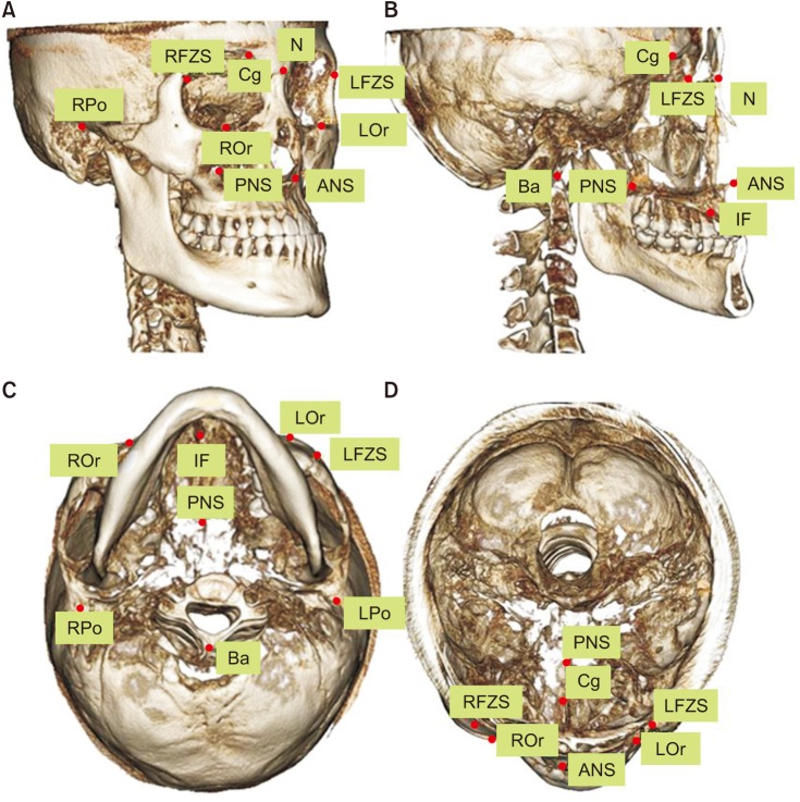
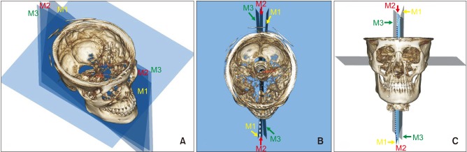
Methods: Forty-three patients (mean age, 23.0 ± 8.20 years) were grouped as follows: group 1 consisted of 10 patients with skeletal Class I and a menton (Me) deviation of < 2 mm; group 2, 11 patients with skeletal Class III and a Me deviation < 2 mm; group 3, nine patients with skeletal Class III and a Me deviation of 2 to less than 4 mm; and group 4, 13 patients with skeletal Class III and an Me deviation ≥ 4 mm. The candidate MSPs were established by three-dimensional (3D) cone beam computed tomography (CBCT) reorientation methods (RMs): (1) the MSP perpendicular to the Frankfort horizontal (FH) plane while passing through the crista galli and basion; (2) the MSP including the nasion, incisive foramen, and basion; (3) the MSP including the nasion, anterior nasal spine, and posterior nasal spine. The mean absolute distances (MADs) to the MSPs were calculated from the coordinates of 1,548 points on 129 CBCT images. The differences in the values of the 3D coordinates among RMs were compared.
Results: The MADs of the three RMs showed significant differences (p < 0.05). Most of the differences in values of the coordinates were not significant among RMs.
Conclusions: Although the differences in distance among the three MSPs were minor, the MSP perpendicular to the FH plane while passing through the crista galli and basion best approximated the true symmetrical MSP.
期刊介绍:
The Korean Journal of Orthodontics (KJO) is an international, open access, peer reviewed journal published in January, March, May, July, September, and November each year. It was first launched in 1970 and, as the official scientific publication of Korean Association of Orthodontists, KJO aims to publish high quality clinical and scientific original research papers in all areas related to orthodontics and dentofacial orthopedics. Specifically, its interest focuses on evidence-based investigations of contemporary diagnostic procedures and treatment techniques, expanding to significant clinical reports of diverse treatment approaches.
The scope of KJO covers all areas of orthodontics and dentofacial orthopedics including successful diagnostic procedures and treatment planning, growth and development of the face and its clinical implications, appliance designs, biomechanics, TMJ disorders and adult treatment. Specifically, its latest interest focuses on skeletal anchorage devices, orthodontic appliance and biomaterials, 3 dimensional imaging techniques utilized for dentofacial diagnosis and treatment planning, and orthognathic surgery to correct skeletal disharmony in association of orthodontic treatment.

 求助内容:
求助内容: 应助结果提醒方式:


