Emanuele F Osimo, Stefan P Brugger, Antonio de Marvao, Toby Pillinger, Thomas Whitehurst, Ben Statton, Marina Quinlan, Alaine Berry, Stuart A Cook, Declan P O'Regan, Oliver D Howes
{"title":"精神分裂症患者心脏结构和功能:心脏磁共振成像研究。","authors":"Emanuele F Osimo, Stefan P Brugger, Antonio de Marvao, Toby Pillinger, Thomas Whitehurst, Ben Statton, Marina Quinlan, Alaine Berry, Stuart A Cook, Declan P O'Regan, Oliver D Howes","doi":"10.1192/bjp.2019.268","DOIUrl":null,"url":null,"abstract":"<p><strong>Background: </strong>Heart disease is the leading cause of death in schizophrenia. However, there has been little research directly examining cardiac function in schizophrenia.</p><p><strong>Aims: </strong>To investigate cardiac structure and function in individuals with schizophrenia using cardiac magnetic resonance imaging (CMR) after excluding medical and metabolic comorbidity.</p><p><strong>Method: </strong>In total, 80 participants underwent CMR to determine biventricular volumes and function and measures of blood pressure, physical activity and glycated haemoglobin levels. Individuals with schizophrenia ('patients') and controls were matched for age, gender, ethnicity and body surface area.</p><p><strong>Results: </strong>Patients had significantly smaller indexed left ventricular (LV) end-diastolic volume (effect size d = -0.82, P = 0.001), LV end-systolic volume (d = -0.58, P = 0.02), LV stroke volume (d = -0.85, P = 0.001), right ventricular (RV) end-diastolic volume (d = -0.79, P = 0.002), RV end-systolic volume (d = -0.58, P = 0.02), and RV stroke volume (d = -0.87, P = 0.001) but unaltered ejection fractions relative to controls. LV concentricity (d = 0.73, P = 0.003) and septal thickness (d = 1.13, P < 0.001) were significantly larger in the patients. Mean concentricity in patients was above the reference range. The findings were largely unchanged after adjusting for smoking and/or exercise levels and were independent of medication dose and duration.</p><p><strong>Conclusions: </strong>Individuals with schizophrenia show evidence of concentric cardiac remodelling compared with healthy controls of a similar age, gender, ethnicity, body surface area and blood pressure, and independent of smoking and activity levels. This could be contributing to the excess cardiovascular mortality observed in schizophrenia. Future studies should investigate the contribution of antipsychotic medication to these changes.</p>","PeriodicalId":520791,"journal":{"name":"The British journal of psychiatry : the journal of mental science","volume":" ","pages":"450-457"},"PeriodicalIF":0.0000,"publicationDate":"2020-08-01","publicationTypes":"Journal Article","fieldsOfStudy":null,"isOpenAccess":false,"openAccessPdf":"https://www.ncbi.nlm.nih.gov/pmc/articles/PMC7511899/pdf/","citationCount":"0","resultStr":"{\"title\":\"Cardiac structure and function in schizophrenia: cardiac magnetic resonance imaging study.\",\"authors\":\"Emanuele F Osimo, Stefan P Brugger, Antonio de Marvao, Toby Pillinger, Thomas Whitehurst, Ben Statton, Marina Quinlan, Alaine Berry, Stuart A Cook, Declan P O'Regan, Oliver D Howes\",\"doi\":\"10.1192/bjp.2019.268\",\"DOIUrl\":null,\"url\":null,\"abstract\":\"<p><strong>Background: </strong>Heart disease is the leading cause of death in schizophrenia. However, there has been little research directly examining cardiac function in schizophrenia.</p><p><strong>Aims: </strong>To investigate cardiac structure and function in individuals with schizophrenia using cardiac magnetic resonance imaging (CMR) after excluding medical and metabolic comorbidity.</p><p><strong>Method: </strong>In total, 80 participants underwent CMR to determine biventricular volumes and function and measures of blood pressure, physical activity and glycated haemoglobin levels. Individuals with schizophrenia ('patients') and controls were matched for age, gender, ethnicity and body surface area.</p><p><strong>Results: </strong>Patients had significantly smaller indexed left ventricular (LV) end-diastolic volume (effect size d = -0.82, P = 0.001), LV end-systolic volume (d = -0.58, P = 0.02), LV stroke volume (d = -0.85, P = 0.001), right ventricular (RV) end-diastolic volume (d = -0.79, P = 0.002), RV end-systolic volume (d = -0.58, P = 0.02), and RV stroke volume (d = -0.87, P = 0.001) but unaltered ejection fractions relative to controls. LV concentricity (d = 0.73, P = 0.003) and septal thickness (d = 1.13, P < 0.001) were significantly larger in the patients. Mean concentricity in patients was above the reference range. The findings were largely unchanged after adjusting for smoking and/or exercise levels and were independent of medication dose and duration.</p><p><strong>Conclusions: </strong>Individuals with schizophrenia show evidence of concentric cardiac remodelling compared with healthy controls of a similar age, gender, ethnicity, body surface area and blood pressure, and independent of smoking and activity levels. This could be contributing to the excess cardiovascular mortality observed in schizophrenia. Future studies should investigate the contribution of antipsychotic medication to these changes.</p>\",\"PeriodicalId\":520791,\"journal\":{\"name\":\"The British journal of psychiatry : the journal of mental science\",\"volume\":\" \",\"pages\":\"450-457\"},\"PeriodicalIF\":0.0000,\"publicationDate\":\"2020-08-01\",\"publicationTypes\":\"Journal Article\",\"fieldsOfStudy\":null,\"isOpenAccess\":false,\"openAccessPdf\":\"https://www.ncbi.nlm.nih.gov/pmc/articles/PMC7511899/pdf/\",\"citationCount\":\"0\",\"resultStr\":null,\"platform\":\"Semanticscholar\",\"paperid\":null,\"PeriodicalName\":\"The British journal of psychiatry : the journal of mental science\",\"FirstCategoryId\":\"1085\",\"ListUrlMain\":\"https://doi.org/10.1192/bjp.2019.268\",\"RegionNum\":0,\"RegionCategory\":null,\"ArticlePicture\":[],\"TitleCN\":null,\"AbstractTextCN\":null,\"PMCID\":null,\"EPubDate\":\"\",\"PubModel\":\"\",\"JCR\":\"\",\"JCRName\":\"\",\"Score\":null,\"Total\":0}","platform":"Semanticscholar","paperid":null,"PeriodicalName":"The British journal of psychiatry : the journal of mental science","FirstCategoryId":"1085","ListUrlMain":"https://doi.org/10.1192/bjp.2019.268","RegionNum":0,"RegionCategory":null,"ArticlePicture":[],"TitleCN":null,"AbstractTextCN":null,"PMCID":null,"EPubDate":"","PubModel":"","JCR":"","JCRName":"","Score":null,"Total":0}
引用次数: 0
摘要
背景:心脏病是精神分裂症患者死亡的主要原因。然而,很少有研究直接检查精神分裂症患者的心脏功能。目的:在排除药物和代谢合并症后,应用心脏磁共振成像(CMR)研究精神分裂症患者的心脏结构和功能。方法:总共有80名参与者接受了CMR,以确定双心室容积和功能,并测量血压、身体活动和糖化血红蛋白水平。精神分裂症患者(“患者”)和对照组在年龄、性别、种族和体表面积方面进行了匹配。结果:患者的左室舒张末期容积(效应值d = -0.82, P = 0.001)、左室收缩末期容积(效应值d = -0.58, P = 0.02)、左室卒中容积(效应值d = -0.85, P = 0.001)、右室舒张末期容积(效应值d = -0.79, P = 0.002)、右室收缩末期容积(效应值d = -0.58, P = 0.02)和右室卒中容积(效应值d = -0.87, P = 0.001)明显小于对照组,但射血分数不变。左室同心度(d = 0.73, P = 0.003)和室间隔厚度(d = 1.13, P < 0.001)显著增大。患者的平均同心度高于参考范围。在调整吸烟和/或运动水平后,研究结果基本上没有变化,并且与药物剂量和持续时间无关。结论:与年龄、性别、种族、体表面积和血压相近的健康对照相比,精神分裂症患者表现出同心心脏重构的证据,且与吸烟和活动水平无关。这可能是导致精神分裂症患者心血管死亡率过高的原因。未来的研究应探讨抗精神病药物对这些变化的作用。
Cardiac structure and function in schizophrenia: cardiac magnetic resonance imaging study.
Background: Heart disease is the leading cause of death in schizophrenia. However, there has been little research directly examining cardiac function in schizophrenia.
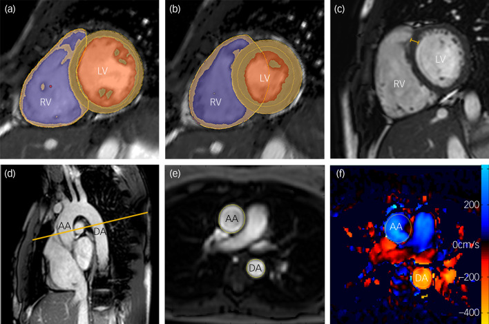
Aims: To investigate cardiac structure and function in individuals with schizophrenia using cardiac magnetic resonance imaging (CMR) after excluding medical and metabolic comorbidity.
Method: In total, 80 participants underwent CMR to determine biventricular volumes and function and measures of blood pressure, physical activity and glycated haemoglobin levels. Individuals with schizophrenia ('patients') and controls were matched for age, gender, ethnicity and body surface area.
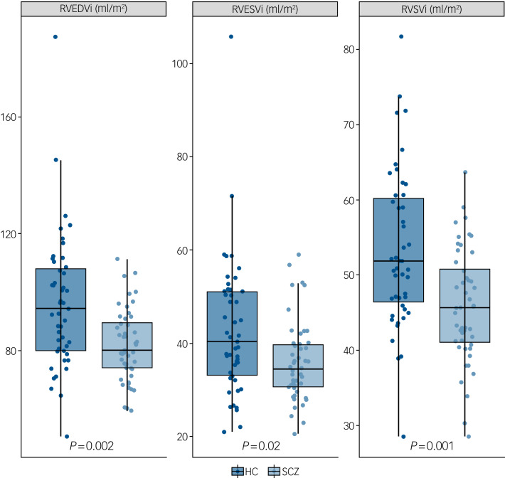
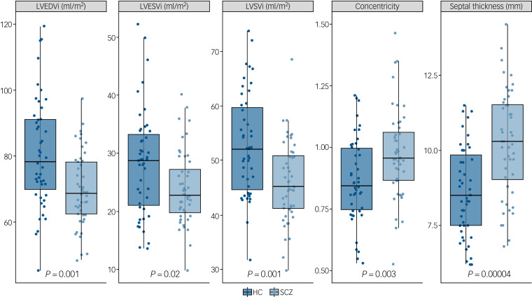
Results: Patients had significantly smaller indexed left ventricular (LV) end-diastolic volume (effect size d = -0.82, P = 0.001), LV end-systolic volume (d = -0.58, P = 0.02), LV stroke volume (d = -0.85, P = 0.001), right ventricular (RV) end-diastolic volume (d = -0.79, P = 0.002), RV end-systolic volume (d = -0.58, P = 0.02), and RV stroke volume (d = -0.87, P = 0.001) but unaltered ejection fractions relative to controls. LV concentricity (d = 0.73, P = 0.003) and septal thickness (d = 1.13, P < 0.001) were significantly larger in the patients. Mean concentricity in patients was above the reference range. The findings were largely unchanged after adjusting for smoking and/or exercise levels and were independent of medication dose and duration.
Conclusions: Individuals with schizophrenia show evidence of concentric cardiac remodelling compared with healthy controls of a similar age, gender, ethnicity, body surface area and blood pressure, and independent of smoking and activity levels. This could be contributing to the excess cardiovascular mortality observed in schizophrenia. Future studies should investigate the contribution of antipsychotic medication to these changes.

 求助内容:
求助内容: 应助结果提醒方式:


