Xiang-Rong Yu, Jun Mao, Wei Tang, Xiang-Ying Meng, Ye Tian, Zhong-Li Du
{"title":"局限于阑尾的低级别阑尾黏液性肿瘤:临床表现和CT表现。","authors":"Xiang-Rong Yu, Jun Mao, Wei Tang, Xiang-Ying Meng, Ye Tian, Zhong-Li Du","doi":"10.1136/jim-2018-000975","DOIUrl":null,"url":null,"abstract":"<p><p>The clinical findings and CT images are investigated in order to fulfill an early preoperative diagnosis and increase awareness of low-grade appendiceal mucinous neoplasm (LAMN) confined to the appendix. 17 cases with histologically proven LAMNs confined to the appendix were included in this study. All patients had received multiphase CT examinations before the surgery. The imaging criteria included shape, size, margin, attenuation, secondary degeneration and internal mass enhancement pattern. In CT images, all cases appeared as oval or tubular cystic masses (average attenuation 20.4±3.6 Hounsfield units), with the longest dimensions ranging from approximately 38 to 106 mm (mean 66.3 mm), and the ratio of length against width was 1.83 in average. The cystic wall was unevenly thickened, with a mean maximal wall thickness of 5.7 mm (>10 mm in 3 cases). The inner capsule wall was rough, and calcification was observed in 3 cases. A few amounts of periappendiceal fat stranding were noted in 2 cases. Mild ring mural enhancement of the cystic wall was seen during the arterial phase, with progressive enhancement during the portal venous phase. In addition, mini enhancing mural nodules was observed in 5 cases. Although preoperative diagnosis of LAMNs confined to the appendix remains challenging, it should be considered when a focal well-defined cystic mass with slightly higher than water attenuation, thickened cystic wall with ring mural enhancement and a characteristic progressive contrast enhancement in CT imaging, especially in older females with non-specific symptoms similar to appendicitis.</p>","PeriodicalId":16112,"journal":{"name":"Journal of Investigative Medicine","volume":"68 1","pages":"75-81"},"PeriodicalIF":2.5000,"publicationDate":"2020-01-01","publicationTypes":"Journal Article","fieldsOfStudy":null,"isOpenAccess":false,"openAccessPdf":"https://sci-hub-pdf.com/10.1136/jim-2018-000975","citationCount":"13","resultStr":"{\"title\":\"Low-grade appendiceal mucinous neoplasms confined to the appendix: clinical manifestations and CT findings.\",\"authors\":\"Xiang-Rong Yu, Jun Mao, Wei Tang, Xiang-Ying Meng, Ye Tian, Zhong-Li Du\",\"doi\":\"10.1136/jim-2018-000975\",\"DOIUrl\":null,\"url\":null,\"abstract\":\"<p><p>The clinical findings and CT images are investigated in order to fulfill an early preoperative diagnosis and increase awareness of low-grade appendiceal mucinous neoplasm (LAMN) confined to the appendix. 17 cases with histologically proven LAMNs confined to the appendix were included in this study. All patients had received multiphase CT examinations before the surgery. The imaging criteria included shape, size, margin, attenuation, secondary degeneration and internal mass enhancement pattern. In CT images, all cases appeared as oval or tubular cystic masses (average attenuation 20.4±3.6 Hounsfield units), with the longest dimensions ranging from approximately 38 to 106 mm (mean 66.3 mm), and the ratio of length against width was 1.83 in average. The cystic wall was unevenly thickened, with a mean maximal wall thickness of 5.7 mm (>10 mm in 3 cases). The inner capsule wall was rough, and calcification was observed in 3 cases. A few amounts of periappendiceal fat stranding were noted in 2 cases. Mild ring mural enhancement of the cystic wall was seen during the arterial phase, with progressive enhancement during the portal venous phase. In addition, mini enhancing mural nodules was observed in 5 cases. Although preoperative diagnosis of LAMNs confined to the appendix remains challenging, it should be considered when a focal well-defined cystic mass with slightly higher than water attenuation, thickened cystic wall with ring mural enhancement and a characteristic progressive contrast enhancement in CT imaging, especially in older females with non-specific symptoms similar to appendicitis.</p>\",\"PeriodicalId\":16112,\"journal\":{\"name\":\"Journal of Investigative Medicine\",\"volume\":\"68 1\",\"pages\":\"75-81\"},\"PeriodicalIF\":2.5000,\"publicationDate\":\"2020-01-01\",\"publicationTypes\":\"Journal Article\",\"fieldsOfStudy\":null,\"isOpenAccess\":false,\"openAccessPdf\":\"https://sci-hub-pdf.com/10.1136/jim-2018-000975\",\"citationCount\":\"13\",\"resultStr\":null,\"platform\":\"Semanticscholar\",\"paperid\":null,\"PeriodicalName\":\"Journal of Investigative Medicine\",\"FirstCategoryId\":\"3\",\"ListUrlMain\":\"https://doi.org/10.1136/jim-2018-000975\",\"RegionNum\":4,\"RegionCategory\":\"医学\",\"ArticlePicture\":[],\"TitleCN\":null,\"AbstractTextCN\":null,\"PMCID\":null,\"EPubDate\":\"2019/7/11 0:00:00\",\"PubModel\":\"Epub\",\"JCR\":\"Q1\",\"JCRName\":\"MEDICINE, GENERAL & INTERNAL\",\"Score\":null,\"Total\":0}","platform":"Semanticscholar","paperid":null,"PeriodicalName":"Journal of Investigative Medicine","FirstCategoryId":"3","ListUrlMain":"https://doi.org/10.1136/jim-2018-000975","RegionNum":4,"RegionCategory":"医学","ArticlePicture":[],"TitleCN":null,"AbstractTextCN":null,"PMCID":null,"EPubDate":"2019/7/11 0:00:00","PubModel":"Epub","JCR":"Q1","JCRName":"MEDICINE, GENERAL & INTERNAL","Score":null,"Total":0}
引用次数: 13
摘要
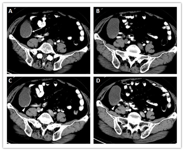
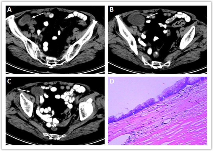
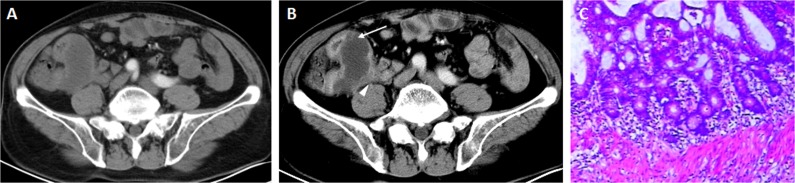
为了实现早期术前诊断和提高对局限于阑尾的低级别阑尾粘液瘤(LAMN)的认识,我们对临床表现和CT图像进行了研究。本研究包括17例组织学证实的局限于阑尾的lamn。所有患者术前均行多期CT检查。影像标准包括形状、大小、边缘、衰减、继发变性和内部肿块增强模式。CT表现均为卵圆形或管状囊性肿块,平均衰减20.4±3.6 Hounsfield单位,最大尺寸约38 ~ 106 mm(平均66.3 mm),长宽比平均1.83。囊壁增厚不均匀,平均最大壁厚5.7 mm (>10 mm 3例)。内囊壁粗糙,3例可见钙化。2例阑尾周围有少量脂肪搁浅。动脉期可见囊壁轻度环形强化,门静脉期渐进式强化。此外,5例患者可见微小强化的壁结节。尽管对于局限于阑尾的LAMNs的术前诊断仍然具有挑战性,但当出现局灶性、界限明确的囊性肿块,其衰减程度略高于水,囊壁增厚伴环状壁增强,CT成像表现为特征性的渐进式增强时,尤其是有类似阑尾炎的非特异性症状的老年女性,应予以考虑。
Low-grade appendiceal mucinous neoplasms confined to the appendix: clinical manifestations and CT findings.
The clinical findings and CT images are investigated in order to fulfill an early preoperative diagnosis and increase awareness of low-grade appendiceal mucinous neoplasm (LAMN) confined to the appendix. 17 cases with histologically proven LAMNs confined to the appendix were included in this study. All patients had received multiphase CT examinations before the surgery. The imaging criteria included shape, size, margin, attenuation, secondary degeneration and internal mass enhancement pattern. In CT images, all cases appeared as oval or tubular cystic masses (average attenuation 20.4±3.6 Hounsfield units), with the longest dimensions ranging from approximately 38 to 106 mm (mean 66.3 mm), and the ratio of length against width was 1.83 in average. The cystic wall was unevenly thickened, with a mean maximal wall thickness of 5.7 mm (>10 mm in 3 cases). The inner capsule wall was rough, and calcification was observed in 3 cases. A few amounts of periappendiceal fat stranding were noted in 2 cases. Mild ring mural enhancement of the cystic wall was seen during the arterial phase, with progressive enhancement during the portal venous phase. In addition, mini enhancing mural nodules was observed in 5 cases. Although preoperative diagnosis of LAMNs confined to the appendix remains challenging, it should be considered when a focal well-defined cystic mass with slightly higher than water attenuation, thickened cystic wall with ring mural enhancement and a characteristic progressive contrast enhancement in CT imaging, especially in older females with non-specific symptoms similar to appendicitis.
期刊介绍:
Journal of Investigative Medicine (JIM) is the official publication of the American Federation for Medical Research. The journal is peer-reviewed and publishes high-quality original articles and reviews in the areas of basic, clinical, and translational medical research.
JIM publishes on all topics and specialty areas that are critical to the conduct of the entire spectrum of biomedical research: from the translation of clinical observations at the bedside, to basic and animal research to clinical research and the implementation of innovative medical care.
 求助内容:
求助内容: 应助结果提醒方式:


