{"title":"正常前列腺及周围结构的3T MR解剖。","authors":"K Sklinda, M Frączek, B Mruk, J Walecki","doi":"10.1155/2019/3040859","DOIUrl":null,"url":null,"abstract":"<p><p>Development on new fast MRI scanners resulted in rising number of prostate examinations. High-spatial resolution of MRI examinations performed on 3T scanners allows recognition of very fine anatomical structures previously not demarcated on performed scans. We present current status of MR imaging in the context of recognition of most important anatomical structures.</p>","PeriodicalId":53309,"journal":{"name":"Advances in Medicine","volume":"2019 ","pages":"3040859"},"PeriodicalIF":0.0000,"publicationDate":"2019-05-28","publicationTypes":"Journal Article","fieldsOfStudy":null,"isOpenAccess":false,"openAccessPdf":"https://sci-hub-pdf.com/10.1155/2019/3040859","citationCount":"12","resultStr":"{\"title\":\"Normal 3T MR Anatomy of the Prostate Gland and Surrounding Structures.\",\"authors\":\"K Sklinda, M Frączek, B Mruk, J Walecki\",\"doi\":\"10.1155/2019/3040859\",\"DOIUrl\":null,\"url\":null,\"abstract\":\"<p><p>Development on new fast MRI scanners resulted in rising number of prostate examinations. High-spatial resolution of MRI examinations performed on 3T scanners allows recognition of very fine anatomical structures previously not demarcated on performed scans. We present current status of MR imaging in the context of recognition of most important anatomical structures.</p>\",\"PeriodicalId\":53309,\"journal\":{\"name\":\"Advances in Medicine\",\"volume\":\"2019 \",\"pages\":\"3040859\"},\"PeriodicalIF\":0.0000,\"publicationDate\":\"2019-05-28\",\"publicationTypes\":\"Journal Article\",\"fieldsOfStudy\":null,\"isOpenAccess\":false,\"openAccessPdf\":\"https://sci-hub-pdf.com/10.1155/2019/3040859\",\"citationCount\":\"12\",\"resultStr\":null,\"platform\":\"Semanticscholar\",\"paperid\":null,\"PeriodicalName\":\"Advances in Medicine\",\"FirstCategoryId\":\"1085\",\"ListUrlMain\":\"https://doi.org/10.1155/2019/3040859\",\"RegionNum\":0,\"RegionCategory\":null,\"ArticlePicture\":[],\"TitleCN\":null,\"AbstractTextCN\":null,\"PMCID\":null,\"EPubDate\":\"2019/1/1 0:00:00\",\"PubModel\":\"eCollection\",\"JCR\":\"\",\"JCRName\":\"\",\"Score\":null,\"Total\":0}","platform":"Semanticscholar","paperid":null,"PeriodicalName":"Advances in Medicine","FirstCategoryId":"1085","ListUrlMain":"https://doi.org/10.1155/2019/3040859","RegionNum":0,"RegionCategory":null,"ArticlePicture":[],"TitleCN":null,"AbstractTextCN":null,"PMCID":null,"EPubDate":"2019/1/1 0:00:00","PubModel":"eCollection","JCR":"","JCRName":"","Score":null,"Total":0}
Normal 3T MR Anatomy of the Prostate Gland and Surrounding Structures.
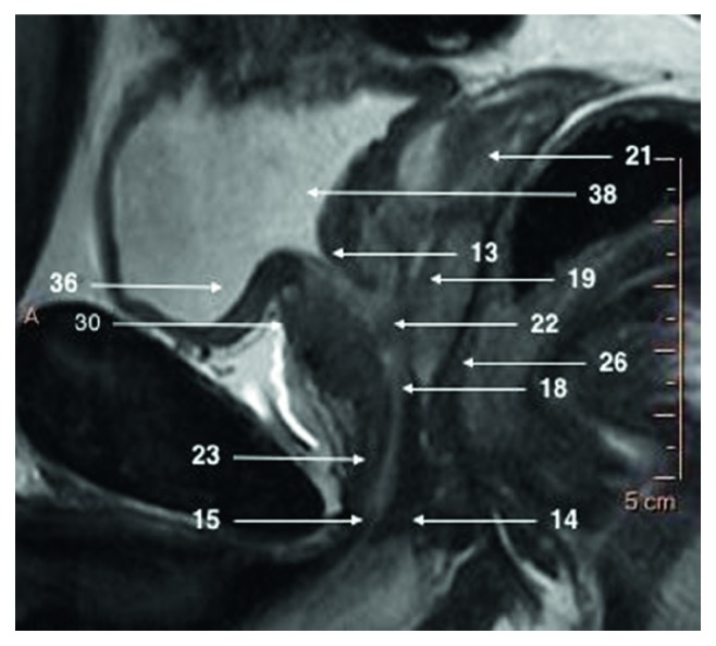
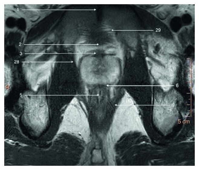
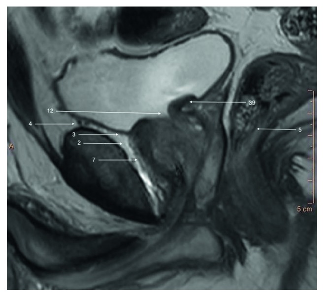
Development on new fast MRI scanners resulted in rising number of prostate examinations. High-spatial resolution of MRI examinations performed on 3T scanners allows recognition of very fine anatomical structures previously not demarcated on performed scans. We present current status of MR imaging in the context of recognition of most important anatomical structures.

 求助内容:
求助内容: 应助结果提醒方式:


