{"title":"睡眠开始过渡期间的脑动力学:脑电图源定位研究","authors":"Antonio Fernandez Guerrero , Peter Achermann","doi":"10.1016/j.nbscr.2018.11.001","DOIUrl":null,"url":null,"abstract":"<div><p>EEG source localization is an essential tool to reveal the cortical sources underlying brain oscillatory activity. We applied LORETA, a technique of EEG source localization, to identify the principal brain areas involved in the process of falling asleep (sleep onset, SO). We localized the contributing brain areas of activity in the classical frequency bands and tracked their temporal evolution (in 2-min intervals from 2 min prior to SO up to 10 min after SO) during a baseline night and subsequent recovery sleep after total sleep deprivation of 40 h.</p><p>Delta activity (0.5–5 Hz) gradually increased both in baseline and recovery sleep, starting in frontal areas and finally involving the entire cortex. This increase was steeper in the recovery condition. The evolution of sigma activity (12–16 Hz) resembled an inverted U-shape in both conditions and the activity was most salient in the parietal cortex. In recovery, sigma activity reached its maximum faster than in baseline, but attained lower levels. Theta activity (5–8 Hz) increased with time in large parts of the occipital lobe (baseline and recovery) and in recovery involved additionally frontal areas. Changes in alpha activity (8–12 Hz) at sleep onset involved large areas of the cortex, whereas activity in the beta range (16–24 Hz) was restricted to small cortical areas. The dynamics in recovery could be considered as a “fast-forward version” of the one in baseline.</p><p>Our results confirm that the process of falling asleep is neither spatially nor temporally a uniform process and that different brain areas might be falling asleep at a different speed potentially reflecting use dependent aspects of sleep regulation.</p></div>","PeriodicalId":37827,"journal":{"name":"Neurobiology of Sleep and Circadian Rhythms","volume":"6 ","pages":"Pages 24-34"},"PeriodicalIF":0.0000,"publicationDate":"2019-01-01","publicationTypes":"Journal Article","fieldsOfStudy":null,"isOpenAccess":false,"openAccessPdf":"https://sci-hub-pdf.com/10.1016/j.nbscr.2018.11.001","citationCount":"24","resultStr":"{\"title\":\"Brain dynamics during the sleep onset transition: An EEG source localization study\",\"authors\":\"Antonio Fernandez Guerrero , Peter Achermann\",\"doi\":\"10.1016/j.nbscr.2018.11.001\",\"DOIUrl\":null,\"url\":null,\"abstract\":\"<div><p>EEG source localization is an essential tool to reveal the cortical sources underlying brain oscillatory activity. We applied LORETA, a technique of EEG source localization, to identify the principal brain areas involved in the process of falling asleep (sleep onset, SO). We localized the contributing brain areas of activity in the classical frequency bands and tracked their temporal evolution (in 2-min intervals from 2 min prior to SO up to 10 min after SO) during a baseline night and subsequent recovery sleep after total sleep deprivation of 40 h.</p><p>Delta activity (0.5–5 Hz) gradually increased both in baseline and recovery sleep, starting in frontal areas and finally involving the entire cortex. This increase was steeper in the recovery condition. The evolution of sigma activity (12–16 Hz) resembled an inverted U-shape in both conditions and the activity was most salient in the parietal cortex. In recovery, sigma activity reached its maximum faster than in baseline, but attained lower levels. Theta activity (5–8 Hz) increased with time in large parts of the occipital lobe (baseline and recovery) and in recovery involved additionally frontal areas. Changes in alpha activity (8–12 Hz) at sleep onset involved large areas of the cortex, whereas activity in the beta range (16–24 Hz) was restricted to small cortical areas. The dynamics in recovery could be considered as a “fast-forward version” of the one in baseline.</p><p>Our results confirm that the process of falling asleep is neither spatially nor temporally a uniform process and that different brain areas might be falling asleep at a different speed potentially reflecting use dependent aspects of sleep regulation.</p></div>\",\"PeriodicalId\":37827,\"journal\":{\"name\":\"Neurobiology of Sleep and Circadian Rhythms\",\"volume\":\"6 \",\"pages\":\"Pages 24-34\"},\"PeriodicalIF\":0.0000,\"publicationDate\":\"2019-01-01\",\"publicationTypes\":\"Journal Article\",\"fieldsOfStudy\":null,\"isOpenAccess\":false,\"openAccessPdf\":\"https://sci-hub-pdf.com/10.1016/j.nbscr.2018.11.001\",\"citationCount\":\"24\",\"resultStr\":null,\"platform\":\"Semanticscholar\",\"paperid\":null,\"PeriodicalName\":\"Neurobiology of Sleep and Circadian Rhythms\",\"FirstCategoryId\":\"1085\",\"ListUrlMain\":\"https://www.sciencedirect.com/science/article/pii/S2451994418300178\",\"RegionNum\":0,\"RegionCategory\":null,\"ArticlePicture\":[],\"TitleCN\":null,\"AbstractTextCN\":null,\"PMCID\":null,\"EPubDate\":\"\",\"PubModel\":\"\",\"JCR\":\"Q2\",\"JCRName\":\"Medicine\",\"Score\":null,\"Total\":0}","platform":"Semanticscholar","paperid":null,"PeriodicalName":"Neurobiology of Sleep and Circadian Rhythms","FirstCategoryId":"1085","ListUrlMain":"https://www.sciencedirect.com/science/article/pii/S2451994418300178","RegionNum":0,"RegionCategory":null,"ArticlePicture":[],"TitleCN":null,"AbstractTextCN":null,"PMCID":null,"EPubDate":"","PubModel":"","JCR":"Q2","JCRName":"Medicine","Score":null,"Total":0}
Brain dynamics during the sleep onset transition: An EEG source localization study
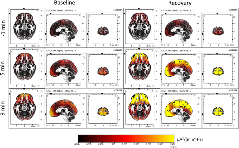
EEG source localization is an essential tool to reveal the cortical sources underlying brain oscillatory activity. We applied LORETA, a technique of EEG source localization, to identify the principal brain areas involved in the process of falling asleep (sleep onset, SO). We localized the contributing brain areas of activity in the classical frequency bands and tracked their temporal evolution (in 2-min intervals from 2 min prior to SO up to 10 min after SO) during a baseline night and subsequent recovery sleep after total sleep deprivation of 40 h.
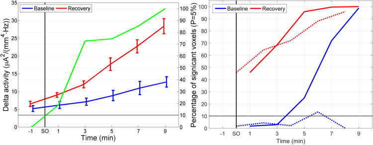
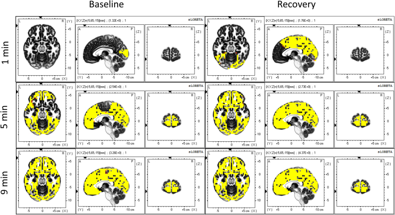
Delta activity (0.5–5 Hz) gradually increased both in baseline and recovery sleep, starting in frontal areas and finally involving the entire cortex. This increase was steeper in the recovery condition. The evolution of sigma activity (12–16 Hz) resembled an inverted U-shape in both conditions and the activity was most salient in the parietal cortex. In recovery, sigma activity reached its maximum faster than in baseline, but attained lower levels. Theta activity (5–8 Hz) increased with time in large parts of the occipital lobe (baseline and recovery) and in recovery involved additionally frontal areas. Changes in alpha activity (8–12 Hz) at sleep onset involved large areas of the cortex, whereas activity in the beta range (16–24 Hz) was restricted to small cortical areas. The dynamics in recovery could be considered as a “fast-forward version” of the one in baseline.
Our results confirm that the process of falling asleep is neither spatially nor temporally a uniform process and that different brain areas might be falling asleep at a different speed potentially reflecting use dependent aspects of sleep regulation.
期刊介绍:
Neurobiology of Sleep and Circadian Rhythms is a multidisciplinary journal for the publication of original research and review articles on basic and translational research into sleep and circadian rhythms. The journal focuses on topics covering the mechanisms of sleep/wake and circadian regulation from molecular to systems level, and on the functional consequences of sleep and circadian disruption. A key aim of the journal is the translation of basic research findings to understand and treat sleep and circadian disorders. Topics include, but are not limited to: Basic and translational research, Molecular mechanisms, Genetics and epigenetics, Inflammation and immunology, Memory and learning, Neurological and neurodegenerative diseases, Neuropsychopharmacology and neuroendocrinology, Behavioral sleep and circadian disorders, Shiftwork, Social jetlag.

 求助内容:
求助内容: 应助结果提醒方式:


