Chadi G Abdallah, Christopher L Averill, Amy E Ramage, Lynnette A Averill, Selin Goktas, Samaneh Nemati, John H Krystal, John D Roache, Patricia A Resick, Stacey Young-McCaughan, Alan L Peterson, Peter Fox
{"title":"美国陆军士兵创伤后应激障碍的显著性网络中断。","authors":"Chadi G Abdallah, Christopher L Averill, Amy E Ramage, Lynnette A Averill, Selin Goktas, Samaneh Nemati, John H Krystal, John D Roache, Patricia A Resick, Stacey Young-McCaughan, Alan L Peterson, Peter Fox","doi":"10.1177/2470547019850467","DOIUrl":null,"url":null,"abstract":"Background Better understanding of the neurobiology of posttraumatic stress disorder (PTSD) may be critical to developing novel, effective therapeutics. Here, we conducted a data-driven investigation using a well-established, graph-based topological measure of nodal strength to determine the extent of functional dysconnectivity in a cohort of active duty U.S. Army soldiers with PTSD compared to controls. Methods A total of 102 participants with (n = 50) or without PTSD (n = 52) completed functional magnetic resonance imaging at rest and during symptom provocation using subject-specific script imagery. Vertex/voxel global brain connectivity with global signal regression (GBCr), a measure of nodal strength, was calculated as the average of its functional connectivity with all other vertices/voxels in the brain gray matter. Results In contrast to resting state, where there were no group differences, we found a significantly higher GBCr during symptom provocation, in PTSD participants compared to controls, in areas within the right hemisphere, including anterior insula, caudal-ventrolateral prefrontal, and rostral-ventrolateral parietal cortices. Overall, these clusters overlapped with the ventral and dorsal salience networks. Post hoc analysis showed increased GBCr in these salience clusters during symptom provocation compared to resting state. In addition, resting-state GBCr in the salience clusters predicted GBCr during symptom provocation in PTSD participants but not in controls. Conclusion In PTSD, increased connectivity within the salience network has been previously hypothesized, based primarily on seed-based connectivity findings. The current results strongly support this hypothesis using whole-brain network measure in a fully data-driven approach. It remains to be seen in future studies whether these identified salience disturbances would normalize following treatment.","PeriodicalId":52315,"journal":{"name":"Chronic Stress","volume":" ","pages":""},"PeriodicalIF":0.0000,"publicationDate":"2019-01-01","publicationTypes":"Journal Article","fieldsOfStudy":null,"isOpenAccess":false,"openAccessPdf":"https://sci-hub-pdf.com/10.1177/2470547019850467","citationCount":"30","resultStr":"{\"title\":\"Salience Network Disruption in U.S. Army Soldiers With Posttraumatic Stress Disorder.\",\"authors\":\"Chadi G Abdallah, Christopher L Averill, Amy E Ramage, Lynnette A Averill, Selin Goktas, Samaneh Nemati, John H Krystal, John D Roache, Patricia A Resick, Stacey Young-McCaughan, Alan L Peterson, Peter Fox\",\"doi\":\"10.1177/2470547019850467\",\"DOIUrl\":null,\"url\":null,\"abstract\":\"Background Better understanding of the neurobiology of posttraumatic stress disorder (PTSD) may be critical to developing novel, effective therapeutics. Here, we conducted a data-driven investigation using a well-established, graph-based topological measure of nodal strength to determine the extent of functional dysconnectivity in a cohort of active duty U.S. Army soldiers with PTSD compared to controls. Methods A total of 102 participants with (n = 50) or without PTSD (n = 52) completed functional magnetic resonance imaging at rest and during symptom provocation using subject-specific script imagery. Vertex/voxel global brain connectivity with global signal regression (GBCr), a measure of nodal strength, was calculated as the average of its functional connectivity with all other vertices/voxels in the brain gray matter. Results In contrast to resting state, where there were no group differences, we found a significantly higher GBCr during symptom provocation, in PTSD participants compared to controls, in areas within the right hemisphere, including anterior insula, caudal-ventrolateral prefrontal, and rostral-ventrolateral parietal cortices. Overall, these clusters overlapped with the ventral and dorsal salience networks. Post hoc analysis showed increased GBCr in these salience clusters during symptom provocation compared to resting state. In addition, resting-state GBCr in the salience clusters predicted GBCr during symptom provocation in PTSD participants but not in controls. Conclusion In PTSD, increased connectivity within the salience network has been previously hypothesized, based primarily on seed-based connectivity findings. The current results strongly support this hypothesis using whole-brain network measure in a fully data-driven approach. It remains to be seen in future studies whether these identified salience disturbances would normalize following treatment.\",\"PeriodicalId\":52315,\"journal\":{\"name\":\"Chronic Stress\",\"volume\":\" \",\"pages\":\"\"},\"PeriodicalIF\":0.0000,\"publicationDate\":\"2019-01-01\",\"publicationTypes\":\"Journal Article\",\"fieldsOfStudy\":null,\"isOpenAccess\":false,\"openAccessPdf\":\"https://sci-hub-pdf.com/10.1177/2470547019850467\",\"citationCount\":\"30\",\"resultStr\":null,\"platform\":\"Semanticscholar\",\"paperid\":null,\"PeriodicalName\":\"Chronic Stress\",\"FirstCategoryId\":\"1085\",\"ListUrlMain\":\"https://doi.org/10.1177/2470547019850467\",\"RegionNum\":0,\"RegionCategory\":null,\"ArticlePicture\":[],\"TitleCN\":null,\"AbstractTextCN\":null,\"PMCID\":null,\"EPubDate\":\"2019/5/15 0:00:00\",\"PubModel\":\"Epub\",\"JCR\":\"Q1\",\"JCRName\":\"Psychology\",\"Score\":null,\"Total\":0}","platform":"Semanticscholar","paperid":null,"PeriodicalName":"Chronic Stress","FirstCategoryId":"1085","ListUrlMain":"https://doi.org/10.1177/2470547019850467","RegionNum":0,"RegionCategory":null,"ArticlePicture":[],"TitleCN":null,"AbstractTextCN":null,"PMCID":null,"EPubDate":"2019/5/15 0:00:00","PubModel":"Epub","JCR":"Q1","JCRName":"Psychology","Score":null,"Total":0}
Salience Network Disruption in U.S. Army Soldiers With Posttraumatic Stress Disorder.
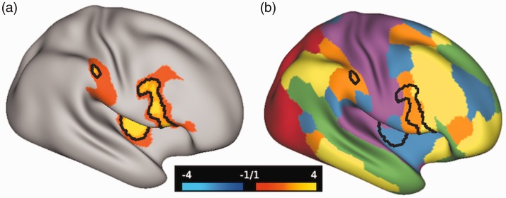
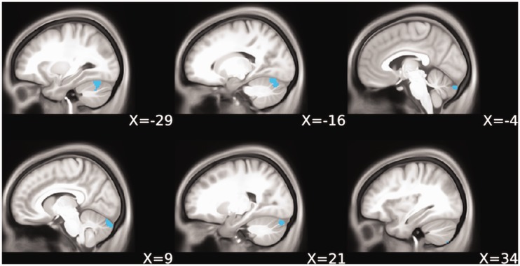
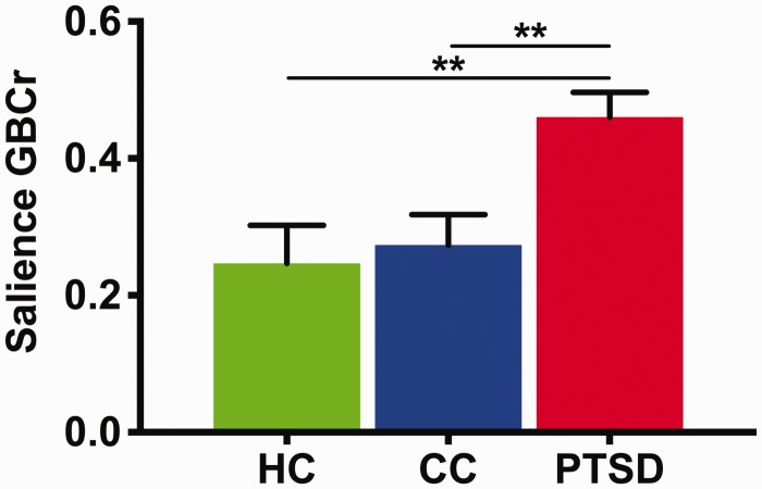
Background Better understanding of the neurobiology of posttraumatic stress disorder (PTSD) may be critical to developing novel, effective therapeutics. Here, we conducted a data-driven investigation using a well-established, graph-based topological measure of nodal strength to determine the extent of functional dysconnectivity in a cohort of active duty U.S. Army soldiers with PTSD compared to controls. Methods A total of 102 participants with (n = 50) or without PTSD (n = 52) completed functional magnetic resonance imaging at rest and during symptom provocation using subject-specific script imagery. Vertex/voxel global brain connectivity with global signal regression (GBCr), a measure of nodal strength, was calculated as the average of its functional connectivity with all other vertices/voxels in the brain gray matter. Results In contrast to resting state, where there were no group differences, we found a significantly higher GBCr during symptom provocation, in PTSD participants compared to controls, in areas within the right hemisphere, including anterior insula, caudal-ventrolateral prefrontal, and rostral-ventrolateral parietal cortices. Overall, these clusters overlapped with the ventral and dorsal salience networks. Post hoc analysis showed increased GBCr in these salience clusters during symptom provocation compared to resting state. In addition, resting-state GBCr in the salience clusters predicted GBCr during symptom provocation in PTSD participants but not in controls. Conclusion In PTSD, increased connectivity within the salience network has been previously hypothesized, based primarily on seed-based connectivity findings. The current results strongly support this hypothesis using whole-brain network measure in a fully data-driven approach. It remains to be seen in future studies whether these identified salience disturbances would normalize following treatment.

 求助内容:
求助内容: 应助结果提醒方式:


