Ji-Lin Li, Shu-Han Lin, Hong-Qiu Chen, Li-Sheng Liang, Xian-Wei Mo, Hao Lai, Jie Zhang, Jing Xu, Bing-Qian Gao, Yan Feng, Yuan Lin
{"title":"结直肠癌卵巢转移患者HER2和EGFR表达的临床意义","authors":"Ji-Lin Li, Shu-Han Lin, Hong-Qiu Chen, Li-Sheng Liang, Xian-Wei Mo, Hao Lai, Jie Zhang, Jing Xu, Bing-Qian Gao, Yan Feng, Yuan Lin","doi":"10.1186/s12907-019-0085-8","DOIUrl":null,"url":null,"abstract":"<p><strong>Background: </strong>EGFR and HER2 overexpression has been reported to play important roles in colorectal cancer (CRC) development and metastasis. Ovarian metastasis is rare yet is one of the most malignant metastases of CRC, but very few studies have focused on its biological features. This study aimed to investigate the expression of EGFR and HER2 in ovarian metastases of CRC and to reveal their clinical significance.</p><p><strong>Methods: </strong>The expression of HER2 and EGFR in both primary tumours and ovarian metastases was analysed by immunohistochemistry (IHC) in 31 CRC patients with ovarian metastases as well as in the primary tumours of 26 CRC patients with non-ovarian metastases. The overall survival time was calculated with a Kaplan-Meier survival curve and compared with a log-rank test.</p><p><strong>Results: </strong>HER2 positivity in primary tumours was significantly higher in patients with ovarian metastases than in those with non-ovarian metastases (54.5% vs. 36.4%, <i>P</i> < 0.05). The EGFR-positive rate in primary lesions was not significantly different between patients with ovarian metastases and those with non-ovarian metastases (63.6% vs. 58.3%, <i>P</i> > 0.05). HER2 expression was not correlated with age, primary tumour site, tumour differentiation, tumour diameter or vascular cancer embolus (<i>P</i> > 0.05). The positive rates of HER2 and EGFR in ovarian metastases were 44.8 and 69.0%, respectively. HER2 expression in ovarian metastases was correlated with peritoneal metastasis and bilateral ovarian metastasis (<i>P</i> < 0.05) but not with age, synchronous or metachronous ovarian metastases and the primary tumour site (<i>P</i> > 0.05). There was no significant correlation between EGFR expression and the clinicopathological features in ovarian metastases (<i>P</i> > 0.05). CRC patients with HER2-positive ovarian metastases showed a shortened overall survival time compared to that of CRC patients with HER2-negative metastases (17.0 ± 5.2 vs. 32.0 ± 8.3 months).</p><p><strong>Conclusion: </strong>Our studies revealed that EGFR and HER2 are highly expressed in the primary tumours and metastases of CRC patients with ovarian metastases. HER2 positivity may be a negative prognostic predictor in patients with ovarian metastases.</p>","PeriodicalId":35804,"journal":{"name":"BMC Clinical Pathology","volume":"19 ","pages":"3"},"PeriodicalIF":0.0000,"publicationDate":"2019-02-28","publicationTypes":"Journal Article","fieldsOfStudy":null,"isOpenAccess":false,"openAccessPdf":"https://sci-hub-pdf.com/10.1186/s12907-019-0085-8","citationCount":"17","resultStr":"{\"title\":\"Clinical significance of HER2 and EGFR expression in colorectal cancer patients with ovarian metastasis.\",\"authors\":\"Ji-Lin Li, Shu-Han Lin, Hong-Qiu Chen, Li-Sheng Liang, Xian-Wei Mo, Hao Lai, Jie Zhang, Jing Xu, Bing-Qian Gao, Yan Feng, Yuan Lin\",\"doi\":\"10.1186/s12907-019-0085-8\",\"DOIUrl\":null,\"url\":null,\"abstract\":\"<p><strong>Background: </strong>EGFR and HER2 overexpression has been reported to play important roles in colorectal cancer (CRC) development and metastasis. Ovarian metastasis is rare yet is one of the most malignant metastases of CRC, but very few studies have focused on its biological features. This study aimed to investigate the expression of EGFR and HER2 in ovarian metastases of CRC and to reveal their clinical significance.</p><p><strong>Methods: </strong>The expression of HER2 and EGFR in both primary tumours and ovarian metastases was analysed by immunohistochemistry (IHC) in 31 CRC patients with ovarian metastases as well as in the primary tumours of 26 CRC patients with non-ovarian metastases. The overall survival time was calculated with a Kaplan-Meier survival curve and compared with a log-rank test.</p><p><strong>Results: </strong>HER2 positivity in primary tumours was significantly higher in patients with ovarian metastases than in those with non-ovarian metastases (54.5% vs. 36.4%, <i>P</i> < 0.05). The EGFR-positive rate in primary lesions was not significantly different between patients with ovarian metastases and those with non-ovarian metastases (63.6% vs. 58.3%, <i>P</i> > 0.05). HER2 expression was not correlated with age, primary tumour site, tumour differentiation, tumour diameter or vascular cancer embolus (<i>P</i> > 0.05). The positive rates of HER2 and EGFR in ovarian metastases were 44.8 and 69.0%, respectively. HER2 expression in ovarian metastases was correlated with peritoneal metastasis and bilateral ovarian metastasis (<i>P</i> < 0.05) but not with age, synchronous or metachronous ovarian metastases and the primary tumour site (<i>P</i> > 0.05). There was no significant correlation between EGFR expression and the clinicopathological features in ovarian metastases (<i>P</i> > 0.05). CRC patients with HER2-positive ovarian metastases showed a shortened overall survival time compared to that of CRC patients with HER2-negative metastases (17.0 ± 5.2 vs. 32.0 ± 8.3 months).</p><p><strong>Conclusion: </strong>Our studies revealed that EGFR and HER2 are highly expressed in the primary tumours and metastases of CRC patients with ovarian metastases. HER2 positivity may be a negative prognostic predictor in patients with ovarian metastases.</p>\",\"PeriodicalId\":35804,\"journal\":{\"name\":\"BMC Clinical Pathology\",\"volume\":\"19 \",\"pages\":\"3\"},\"PeriodicalIF\":0.0000,\"publicationDate\":\"2019-02-28\",\"publicationTypes\":\"Journal Article\",\"fieldsOfStudy\":null,\"isOpenAccess\":false,\"openAccessPdf\":\"https://sci-hub-pdf.com/10.1186/s12907-019-0085-8\",\"citationCount\":\"17\",\"resultStr\":null,\"platform\":\"Semanticscholar\",\"paperid\":null,\"PeriodicalName\":\"BMC Clinical Pathology\",\"FirstCategoryId\":\"1085\",\"ListUrlMain\":\"https://doi.org/10.1186/s12907-019-0085-8\",\"RegionNum\":0,\"RegionCategory\":null,\"ArticlePicture\":[],\"TitleCN\":null,\"AbstractTextCN\":null,\"PMCID\":null,\"EPubDate\":\"2019/1/1 0:00:00\",\"PubModel\":\"eCollection\",\"JCR\":\"Q2\",\"JCRName\":\"Medicine\",\"Score\":null,\"Total\":0}","platform":"Semanticscholar","paperid":null,"PeriodicalName":"BMC Clinical Pathology","FirstCategoryId":"1085","ListUrlMain":"https://doi.org/10.1186/s12907-019-0085-8","RegionNum":0,"RegionCategory":null,"ArticlePicture":[],"TitleCN":null,"AbstractTextCN":null,"PMCID":null,"EPubDate":"2019/1/1 0:00:00","PubModel":"eCollection","JCR":"Q2","JCRName":"Medicine","Score":null,"Total":0}
引用次数: 17
摘要
背景:EGFR和HER2过表达在结直肠癌(CRC)的发展和转移中起重要作用。卵巢转移是结直肠癌最罕见的恶性转移之一,但对其生物学特征的研究很少。本研究旨在探讨EGFR和HER2在结直肠癌卵巢转移灶中的表达及其临床意义。方法:应用免疫组化(IHC)方法分析31例结直肠癌卵巢转移患者原发肿瘤和卵巢转移灶中HER2和EGFR的表达,以及26例结直肠癌原发肿瘤中非卵巢转移灶的表达。用Kaplan-Meier生存曲线计算总生存时间,并与log-rank检验进行比较。结果:原发性肿瘤中卵巢转移患者HER2阳性表达明显高于非卵巢转移患者(54.5% vs. 36.4%, P P > 0.05)。HER2表达与年龄、原发肿瘤部位、肿瘤分化程度、肿瘤直径、血管癌栓子无关(P > 0.05)。卵巢转移灶中HER2和EGFR的阳性率分别为44.8%和69.0%。卵巢转移灶中HER2表达与腹膜转移及双侧卵巢转移相关(P > 0.05)。EGFR表达与卵巢转移灶的临床病理特征无显著相关性(P > 0.05)。her2卵巢转移阳性的结直肠癌患者的总生存时间比her2卵巢转移阴性的结直肠癌患者短(17.0±5.2 vs. 32.0±8.3个月)。结论:我们的研究表明,EGFR和HER2在结直肠癌卵巢转移患者的原发肿瘤和转移瘤中高表达。HER2阳性可能是卵巢转移患者的阴性预后预测因子。
Clinical significance of HER2 and EGFR expression in colorectal cancer patients with ovarian metastasis.
Background: EGFR and HER2 overexpression has been reported to play important roles in colorectal cancer (CRC) development and metastasis. Ovarian metastasis is rare yet is one of the most malignant metastases of CRC, but very few studies have focused on its biological features. This study aimed to investigate the expression of EGFR and HER2 in ovarian metastases of CRC and to reveal their clinical significance.
Methods: The expression of HER2 and EGFR in both primary tumours and ovarian metastases was analysed by immunohistochemistry (IHC) in 31 CRC patients with ovarian metastases as well as in the primary tumours of 26 CRC patients with non-ovarian metastases. The overall survival time was calculated with a Kaplan-Meier survival curve and compared with a log-rank test.
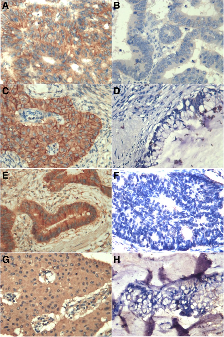
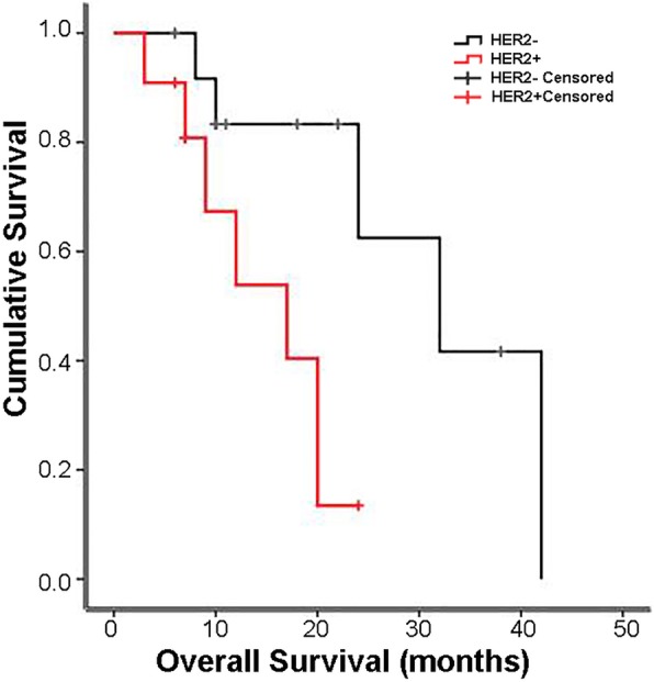
Results: HER2 positivity in primary tumours was significantly higher in patients with ovarian metastases than in those with non-ovarian metastases (54.5% vs. 36.4%, P < 0.05). The EGFR-positive rate in primary lesions was not significantly different between patients with ovarian metastases and those with non-ovarian metastases (63.6% vs. 58.3%, P > 0.05). HER2 expression was not correlated with age, primary tumour site, tumour differentiation, tumour diameter or vascular cancer embolus (P > 0.05). The positive rates of HER2 and EGFR in ovarian metastases were 44.8 and 69.0%, respectively. HER2 expression in ovarian metastases was correlated with peritoneal metastasis and bilateral ovarian metastasis (P < 0.05) but not with age, synchronous or metachronous ovarian metastases and the primary tumour site (P > 0.05). There was no significant correlation between EGFR expression and the clinicopathological features in ovarian metastases (P > 0.05). CRC patients with HER2-positive ovarian metastases showed a shortened overall survival time compared to that of CRC patients with HER2-negative metastases (17.0 ± 5.2 vs. 32.0 ± 8.3 months).
Conclusion: Our studies revealed that EGFR and HER2 are highly expressed in the primary tumours and metastases of CRC patients with ovarian metastases. HER2 positivity may be a negative prognostic predictor in patients with ovarian metastases.
期刊介绍:
BMC Clinical Pathology is an open access journal publishing original peer-reviewed research articles in all aspects of histopathology, haematology, clinical biochemistry, and medical microbiology (including virology, parasitology, and infection control). BMC Clinical Pathology (ISSN 1472-6890) is indexed/tracked/covered by PubMed, CAS, EMBASE, Scopus and Google Scholar.

 求助内容:
求助内容: 应助结果提醒方式:


