{"title":"一种内侧前额皮质细胞类型,对快速抗抑郁反应是必要和充分的。","authors":"Brendan D Hare, Ronald S Duman","doi":"10.1177/2470547019841358","DOIUrl":null,"url":null,"abstract":"Commentary on: Hare BD, Shinohara R, Liu RJ, Pothula S, DiLeone RJ and Duman RS. Optogenetic stimulation of medial prefrontal cortex Drd1 neurons produces rapid and long-lasting antidepressant effects. Nature communications. 2019; 10: 223. The discovery of ketamine’s antidepressant effects has generated great excitement due to the rapid and sustained time course (within hours, lasting up to seven days), efficacy in treatment resistant individuals, as well as a pharmacological profile that is distinct from traditional antidepressants. Efforts have been ongoing to identify the molecular, cellular, and circuit mechanisms underlying the actions of ketamine. Regarding circuitry, dysfunction of the medial prefrontal cortex (mPFC) subregions, including the subgenual and anterior cingulate, have been implicated in depression, and preclinical models have been utilized to study the role of mPFC in depressionlike behaviors. Ketamine is an NMDA antagonist yet it produces a burst of glutamate in the mPFC following administration. Preclinical studies have demonstrated that mPFC activity following ketamine administration is necessary for ketamine’s rapid antidepressant effects, and that activation of principal neurons in the mPFC can produce persistent antidepressant responses. These findings indicate that neurons and circuits within the mPFC may represent key targets in depression. Through interaction with downstream targets, the mPFC plays a role in the generation of numerous behaviors as well as the response to stress. This diversity of action is mediated by principal neurons projecting to numerous downstream targets. Notably, mPFC principal neurons are heterogeneous and have been characterized based on their projection targets, dendritic morphology, and response to neuromodulators. Although the burst of glutamate following ketamine administration may excite many types of mPFC pyramidal neurons indiscriminately, we hypothesized that the antidepressant response may be carried by a discriminable population. To probe this hypothesis, we utilized Cre-dependent expression of neuronal effectors in mouse lines carrying the Cre transgene in neurons expressing the D1 dopamine receptor (Drd1-Cre) or the D2 dopamine receptor (Drd2Cre). This approach had previously been demonstrated to allow cell-type-specific access to distinct populations of mPFC pyramidal cells. We observed that utilizing a channelrhodopsin vector to allow optogenetic stimulation of Drd1 containing cells produced a rapid (observable 24 h after stimulation) and sustained (observable seven days after stimulation) antidepressant response (Figure 1). These effects are similar to ketamine and indicate that the circuit function responsible for depressionlike behavior was rapidly and persistently altered by the prior period of stimulation. In contrast, stimulation of the Drd2 cell type did not produce an antidepressant response. Additional experiments produced convergent evidence for the importance of the Drd1 cell type to the antidepressant response. Chemogenetic inhibition of mPFC Drd1 expressing cells blocked the antidepressant response to ketamine, and D1 antagonist administration into the mPFC when ketamine was administered also eliminated the ketamine response. Notably, using a","PeriodicalId":52315,"journal":{"name":"Chronic Stress","volume":" ","pages":""},"PeriodicalIF":0.0000,"publicationDate":"2019-01-01","publicationTypes":"Journal Article","fieldsOfStudy":null,"isOpenAccess":false,"openAccessPdf":"https://sci-hub-pdf.com/10.1177/2470547019841358","citationCount":"0","resultStr":"{\"title\":\"A medial prefrontal cortex cell type necessary and sufficient for a rapid antidepressant response.\",\"authors\":\"Brendan D Hare, Ronald S Duman\",\"doi\":\"10.1177/2470547019841358\",\"DOIUrl\":null,\"url\":null,\"abstract\":\"Commentary on: Hare BD, Shinohara R, Liu RJ, Pothula S, DiLeone RJ and Duman RS. Optogenetic stimulation of medial prefrontal cortex Drd1 neurons produces rapid and long-lasting antidepressant effects. Nature communications. 2019; 10: 223. The discovery of ketamine’s antidepressant effects has generated great excitement due to the rapid and sustained time course (within hours, lasting up to seven days), efficacy in treatment resistant individuals, as well as a pharmacological profile that is distinct from traditional antidepressants. Efforts have been ongoing to identify the molecular, cellular, and circuit mechanisms underlying the actions of ketamine. Regarding circuitry, dysfunction of the medial prefrontal cortex (mPFC) subregions, including the subgenual and anterior cingulate, have been implicated in depression, and preclinical models have been utilized to study the role of mPFC in depressionlike behaviors. Ketamine is an NMDA antagonist yet it produces a burst of glutamate in the mPFC following administration. Preclinical studies have demonstrated that mPFC activity following ketamine administration is necessary for ketamine’s rapid antidepressant effects, and that activation of principal neurons in the mPFC can produce persistent antidepressant responses. These findings indicate that neurons and circuits within the mPFC may represent key targets in depression. Through interaction with downstream targets, the mPFC plays a role in the generation of numerous behaviors as well as the response to stress. This diversity of action is mediated by principal neurons projecting to numerous downstream targets. Notably, mPFC principal neurons are heterogeneous and have been characterized based on their projection targets, dendritic morphology, and response to neuromodulators. Although the burst of glutamate following ketamine administration may excite many types of mPFC pyramidal neurons indiscriminately, we hypothesized that the antidepressant response may be carried by a discriminable population. To probe this hypothesis, we utilized Cre-dependent expression of neuronal effectors in mouse lines carrying the Cre transgene in neurons expressing the D1 dopamine receptor (Drd1-Cre) or the D2 dopamine receptor (Drd2Cre). This approach had previously been demonstrated to allow cell-type-specific access to distinct populations of mPFC pyramidal cells. We observed that utilizing a channelrhodopsin vector to allow optogenetic stimulation of Drd1 containing cells produced a rapid (observable 24 h after stimulation) and sustained (observable seven days after stimulation) antidepressant response (Figure 1). These effects are similar to ketamine and indicate that the circuit function responsible for depressionlike behavior was rapidly and persistently altered by the prior period of stimulation. In contrast, stimulation of the Drd2 cell type did not produce an antidepressant response. Additional experiments produced convergent evidence for the importance of the Drd1 cell type to the antidepressant response. Chemogenetic inhibition of mPFC Drd1 expressing cells blocked the antidepressant response to ketamine, and D1 antagonist administration into the mPFC when ketamine was administered also eliminated the ketamine response. Notably, using a\",\"PeriodicalId\":52315,\"journal\":{\"name\":\"Chronic Stress\",\"volume\":\" \",\"pages\":\"\"},\"PeriodicalIF\":0.0000,\"publicationDate\":\"2019-01-01\",\"publicationTypes\":\"Journal Article\",\"fieldsOfStudy\":null,\"isOpenAccess\":false,\"openAccessPdf\":\"https://sci-hub-pdf.com/10.1177/2470547019841358\",\"citationCount\":\"0\",\"resultStr\":null,\"platform\":\"Semanticscholar\",\"paperid\":null,\"PeriodicalName\":\"Chronic Stress\",\"FirstCategoryId\":\"1085\",\"ListUrlMain\":\"https://doi.org/10.1177/2470547019841358\",\"RegionNum\":0,\"RegionCategory\":null,\"ArticlePicture\":[],\"TitleCN\":null,\"AbstractTextCN\":null,\"PMCID\":null,\"EPubDate\":\"2019/4/15 0:00:00\",\"PubModel\":\"Epub\",\"JCR\":\"Q1\",\"JCRName\":\"Psychology\",\"Score\":null,\"Total\":0}","platform":"Semanticscholar","paperid":null,"PeriodicalName":"Chronic Stress","FirstCategoryId":"1085","ListUrlMain":"https://doi.org/10.1177/2470547019841358","RegionNum":0,"RegionCategory":null,"ArticlePicture":[],"TitleCN":null,"AbstractTextCN":null,"PMCID":null,"EPubDate":"2019/4/15 0:00:00","PubModel":"Epub","JCR":"Q1","JCRName":"Psychology","Score":null,"Total":0}
A medial prefrontal cortex cell type necessary and sufficient for a rapid antidepressant response.
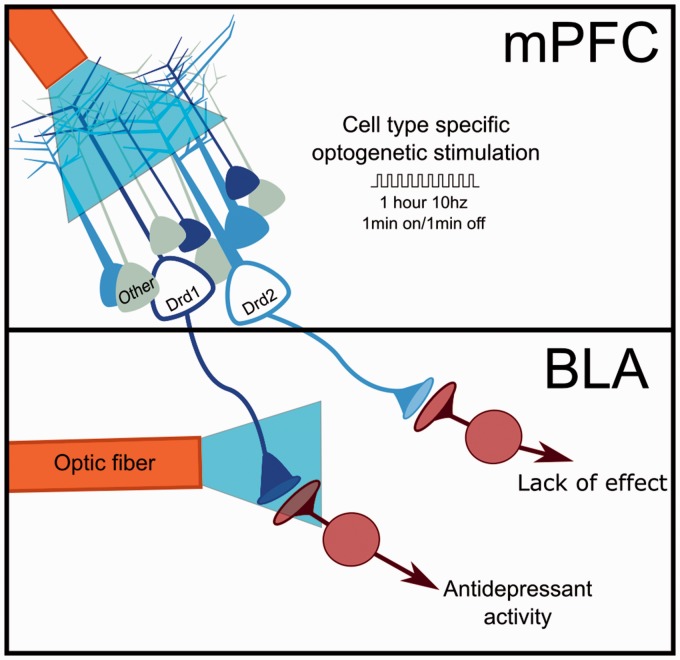
Commentary on: Hare BD, Shinohara R, Liu RJ, Pothula S, DiLeone RJ and Duman RS. Optogenetic stimulation of medial prefrontal cortex Drd1 neurons produces rapid and long-lasting antidepressant effects. Nature communications. 2019; 10: 223. The discovery of ketamine’s antidepressant effects has generated great excitement due to the rapid and sustained time course (within hours, lasting up to seven days), efficacy in treatment resistant individuals, as well as a pharmacological profile that is distinct from traditional antidepressants. Efforts have been ongoing to identify the molecular, cellular, and circuit mechanisms underlying the actions of ketamine. Regarding circuitry, dysfunction of the medial prefrontal cortex (mPFC) subregions, including the subgenual and anterior cingulate, have been implicated in depression, and preclinical models have been utilized to study the role of mPFC in depressionlike behaviors. Ketamine is an NMDA antagonist yet it produces a burst of glutamate in the mPFC following administration. Preclinical studies have demonstrated that mPFC activity following ketamine administration is necessary for ketamine’s rapid antidepressant effects, and that activation of principal neurons in the mPFC can produce persistent antidepressant responses. These findings indicate that neurons and circuits within the mPFC may represent key targets in depression. Through interaction with downstream targets, the mPFC plays a role in the generation of numerous behaviors as well as the response to stress. This diversity of action is mediated by principal neurons projecting to numerous downstream targets. Notably, mPFC principal neurons are heterogeneous and have been characterized based on their projection targets, dendritic morphology, and response to neuromodulators. Although the burst of glutamate following ketamine administration may excite many types of mPFC pyramidal neurons indiscriminately, we hypothesized that the antidepressant response may be carried by a discriminable population. To probe this hypothesis, we utilized Cre-dependent expression of neuronal effectors in mouse lines carrying the Cre transgene in neurons expressing the D1 dopamine receptor (Drd1-Cre) or the D2 dopamine receptor (Drd2Cre). This approach had previously been demonstrated to allow cell-type-specific access to distinct populations of mPFC pyramidal cells. We observed that utilizing a channelrhodopsin vector to allow optogenetic stimulation of Drd1 containing cells produced a rapid (observable 24 h after stimulation) and sustained (observable seven days after stimulation) antidepressant response (Figure 1). These effects are similar to ketamine and indicate that the circuit function responsible for depressionlike behavior was rapidly and persistently altered by the prior period of stimulation. In contrast, stimulation of the Drd2 cell type did not produce an antidepressant response. Additional experiments produced convergent evidence for the importance of the Drd1 cell type to the antidepressant response. Chemogenetic inhibition of mPFC Drd1 expressing cells blocked the antidepressant response to ketamine, and D1 antagonist administration into the mPFC when ketamine was administered also eliminated the ketamine response. Notably, using a

 求助内容:
求助内容: 应助结果提醒方式:


