{"title":"以硬化区为主的肝硬化性血管瘤与胆道囊腺癌相似。","authors":"Hiroyuki Sugo, Yuki Sekine, Shozo Miyano, Ikuo Watanobe, Michio Machida, Kuniaki Kojima, Hironao Okubo, Ayako Ura, Kanako Ogura, Toshiharu Matsumoto","doi":"10.1155/2018/7353170","DOIUrl":null,"url":null,"abstract":"<p><p>We report here an extremely rare case of hepatic sclerosing hemangioma mimicking a biliary cystadenocarcinoma. A previously healthy 39-year-old woman was referred to our hospital because of a large tumor in the liver. Abdominal computed tomography revealed early peripheral ring enhancement in the arterial phase and slight internal heterogeneous enhancement in the delayed phase. Magnetic resonance imaging revealed a tumor with low intensity in the T1-weighted image and very high intensity in the fat-saturated T2-weighted image. The patient underwent hepatectomy for a possible malignant liver tumor. Grossly, the tumor appeared as a white, solid, and cystic mass (weighted 1.1 kg and measured 170×100×80 mm) that was elastic, soft, and homogeneous with a yellowish area. Histological examination showed that the tumor mostly consisted of fibrotic areas with hyalinization. The typical histology of cavernous hemangioma was confirmed in part, and the tumor was diagnosed as a sclerosing hemangioma with predominancy of the sclerosed area. A review of 20 cases reported previously revealed that only 2 (10%) patients were diagnosed as having sclerosing hemangioma preoperatively.</p>","PeriodicalId":30295,"journal":{"name":"Case Reports in Hepatology","volume":"2018 ","pages":"7353170"},"PeriodicalIF":0.0000,"publicationDate":"2018-10-04","publicationTypes":"Journal Article","fieldsOfStudy":null,"isOpenAccess":false,"openAccessPdf":"https://sci-hub-pdf.com/10.1155/2018/7353170","citationCount":"9","resultStr":"{\"title\":\"Hepatic Sclerosing Hemangioma with Predominance of the Sclerosed Area Mimicking a Biliary Cystadenocarcinoma.\",\"authors\":\"Hiroyuki Sugo, Yuki Sekine, Shozo Miyano, Ikuo Watanobe, Michio Machida, Kuniaki Kojima, Hironao Okubo, Ayako Ura, Kanako Ogura, Toshiharu Matsumoto\",\"doi\":\"10.1155/2018/7353170\",\"DOIUrl\":null,\"url\":null,\"abstract\":\"<p><p>We report here an extremely rare case of hepatic sclerosing hemangioma mimicking a biliary cystadenocarcinoma. A previously healthy 39-year-old woman was referred to our hospital because of a large tumor in the liver. Abdominal computed tomography revealed early peripheral ring enhancement in the arterial phase and slight internal heterogeneous enhancement in the delayed phase. Magnetic resonance imaging revealed a tumor with low intensity in the T1-weighted image and very high intensity in the fat-saturated T2-weighted image. The patient underwent hepatectomy for a possible malignant liver tumor. Grossly, the tumor appeared as a white, solid, and cystic mass (weighted 1.1 kg and measured 170×100×80 mm) that was elastic, soft, and homogeneous with a yellowish area. Histological examination showed that the tumor mostly consisted of fibrotic areas with hyalinization. The typical histology of cavernous hemangioma was confirmed in part, and the tumor was diagnosed as a sclerosing hemangioma with predominancy of the sclerosed area. A review of 20 cases reported previously revealed that only 2 (10%) patients were diagnosed as having sclerosing hemangioma preoperatively.</p>\",\"PeriodicalId\":30295,\"journal\":{\"name\":\"Case Reports in Hepatology\",\"volume\":\"2018 \",\"pages\":\"7353170\"},\"PeriodicalIF\":0.0000,\"publicationDate\":\"2018-10-04\",\"publicationTypes\":\"Journal Article\",\"fieldsOfStudy\":null,\"isOpenAccess\":false,\"openAccessPdf\":\"https://sci-hub-pdf.com/10.1155/2018/7353170\",\"citationCount\":\"9\",\"resultStr\":null,\"platform\":\"Semanticscholar\",\"paperid\":null,\"PeriodicalName\":\"Case Reports in Hepatology\",\"FirstCategoryId\":\"1085\",\"ListUrlMain\":\"https://doi.org/10.1155/2018/7353170\",\"RegionNum\":0,\"RegionCategory\":null,\"ArticlePicture\":[],\"TitleCN\":null,\"AbstractTextCN\":null,\"PMCID\":null,\"EPubDate\":\"2018/1/1 0:00:00\",\"PubModel\":\"eCollection\",\"JCR\":\"\",\"JCRName\":\"\",\"Score\":null,\"Total\":0}","platform":"Semanticscholar","paperid":null,"PeriodicalName":"Case Reports in Hepatology","FirstCategoryId":"1085","ListUrlMain":"https://doi.org/10.1155/2018/7353170","RegionNum":0,"RegionCategory":null,"ArticlePicture":[],"TitleCN":null,"AbstractTextCN":null,"PMCID":null,"EPubDate":"2018/1/1 0:00:00","PubModel":"eCollection","JCR":"","JCRName":"","Score":null,"Total":0}
Hepatic Sclerosing Hemangioma with Predominance of the Sclerosed Area Mimicking a Biliary Cystadenocarcinoma.
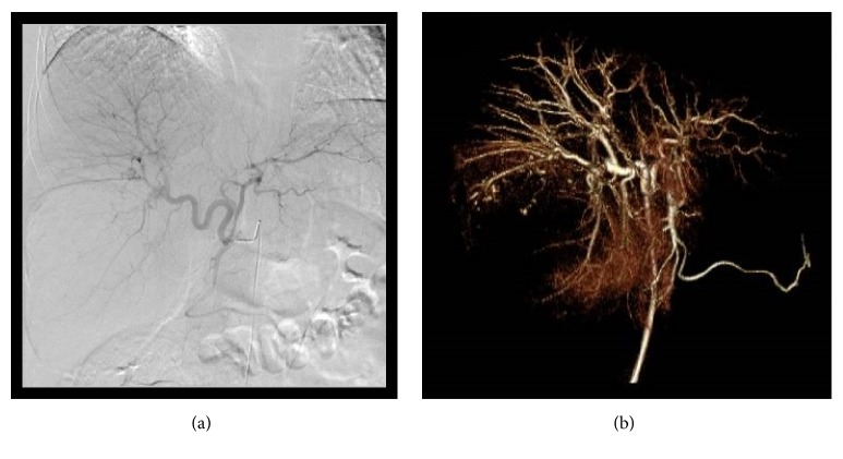
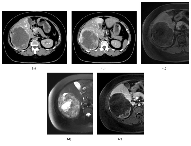
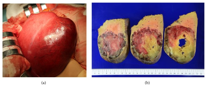
We report here an extremely rare case of hepatic sclerosing hemangioma mimicking a biliary cystadenocarcinoma. A previously healthy 39-year-old woman was referred to our hospital because of a large tumor in the liver. Abdominal computed tomography revealed early peripheral ring enhancement in the arterial phase and slight internal heterogeneous enhancement in the delayed phase. Magnetic resonance imaging revealed a tumor with low intensity in the T1-weighted image and very high intensity in the fat-saturated T2-weighted image. The patient underwent hepatectomy for a possible malignant liver tumor. Grossly, the tumor appeared as a white, solid, and cystic mass (weighted 1.1 kg and measured 170×100×80 mm) that was elastic, soft, and homogeneous with a yellowish area. Histological examination showed that the tumor mostly consisted of fibrotic areas with hyalinization. The typical histology of cavernous hemangioma was confirmed in part, and the tumor was diagnosed as a sclerosing hemangioma with predominancy of the sclerosed area. A review of 20 cases reported previously revealed that only 2 (10%) patients were diagnosed as having sclerosing hemangioma preoperatively.

 求助内容:
求助内容: 应助结果提醒方式:


