Mohammed Q Qutaish, Zhuxian Zhou, David Prabhu, Yiqiao Liu, Mallory R Busso, Donna Izadnegahdar, Madhusudhana Gargesha, Hong Lu, Zheng-Rong Lu, David L Wilson
{"title":"冷冻成像及微转移瘤分子磁共振成像分析软件平台。","authors":"Mohammed Q Qutaish, Zhuxian Zhou, David Prabhu, Yiqiao Liu, Mallory R Busso, Donna Izadnegahdar, Madhusudhana Gargesha, Hong Lu, Zheng-Rong Lu, David L Wilson","doi":"10.1155/2018/9780349","DOIUrl":null,"url":null,"abstract":"<p><p>We created and evaluated a preclinical, multimodality imaging, and software platform to assess molecular imaging of small metastases. This included experimental methods (e.g., GFP-labeled tumor and high resolution multispectral cryo-imaging), nonrigid image registration, and interactive visualization of imaging agent targeting. We describe technological details earlier applied to GFP-labeled metastatic tumor targeting by molecular MR (CREKA-Gd) and red fluorescent (CREKA-Cy5) imaging agents. Optimized nonrigid cryo-MRI registration enabled nonambiguous association of MR signals to GFP tumors. Interactive visualization of out-of-RAM volumetric image data allowed one to zoom to a GFP-labeled micrometastasis, determine its anatomical location from color cryo-images, and establish the presence/absence of targeted CREKA-Gd and CREKA-Cy5. In a mouse with >160 GFP-labeled tumors, we determined that in the MR images every tumor in the lung >0.3 mm<sup>2</sup> had visible signal and that some metastases as small as 0.1 mm<sup>2</sup> were also visible. More tumors were visible in CREKA-Cy5 than in CREKA-Gd MRI. Tape transfer method and nonrigid registration allowed accurate (<11 <i>μ</i>m error) registration of whole mouse histology to corresponding cryo-images. Histology showed inflammation and necrotic regions not labeled by imaging agents. This mouse-to-cells multiscale and multimodality platform should uniquely enable more informative and accurate studies of metastatic cancer imaging and therapy.</p>","PeriodicalId":47063,"journal":{"name":"International Journal of Biomedical Imaging","volume":"2018 ","pages":"9780349"},"PeriodicalIF":1.3000,"publicationDate":"2018-04-01","publicationTypes":"Journal Article","fieldsOfStudy":null,"isOpenAccess":false,"openAccessPdf":"https://sci-hub-pdf.com/10.1155/2018/9780349","citationCount":"12","resultStr":"{\"title\":\"Cryo-Imaging and Software Platform for Analysis of Molecular MR Imaging of Micrometastases.\",\"authors\":\"Mohammed Q Qutaish, Zhuxian Zhou, David Prabhu, Yiqiao Liu, Mallory R Busso, Donna Izadnegahdar, Madhusudhana Gargesha, Hong Lu, Zheng-Rong Lu, David L Wilson\",\"doi\":\"10.1155/2018/9780349\",\"DOIUrl\":null,\"url\":null,\"abstract\":\"<p><p>We created and evaluated a preclinical, multimodality imaging, and software platform to assess molecular imaging of small metastases. This included experimental methods (e.g., GFP-labeled tumor and high resolution multispectral cryo-imaging), nonrigid image registration, and interactive visualization of imaging agent targeting. We describe technological details earlier applied to GFP-labeled metastatic tumor targeting by molecular MR (CREKA-Gd) and red fluorescent (CREKA-Cy5) imaging agents. Optimized nonrigid cryo-MRI registration enabled nonambiguous association of MR signals to GFP tumors. Interactive visualization of out-of-RAM volumetric image data allowed one to zoom to a GFP-labeled micrometastasis, determine its anatomical location from color cryo-images, and establish the presence/absence of targeted CREKA-Gd and CREKA-Cy5. In a mouse with >160 GFP-labeled tumors, we determined that in the MR images every tumor in the lung >0.3 mm<sup>2</sup> had visible signal and that some metastases as small as 0.1 mm<sup>2</sup> were also visible. More tumors were visible in CREKA-Cy5 than in CREKA-Gd MRI. Tape transfer method and nonrigid registration allowed accurate (<11 <i>μ</i>m error) registration of whole mouse histology to corresponding cryo-images. Histology showed inflammation and necrotic regions not labeled by imaging agents. This mouse-to-cells multiscale and multimodality platform should uniquely enable more informative and accurate studies of metastatic cancer imaging and therapy.</p>\",\"PeriodicalId\":47063,\"journal\":{\"name\":\"International Journal of Biomedical Imaging\",\"volume\":\"2018 \",\"pages\":\"9780349\"},\"PeriodicalIF\":1.3000,\"publicationDate\":\"2018-04-01\",\"publicationTypes\":\"Journal Article\",\"fieldsOfStudy\":null,\"isOpenAccess\":false,\"openAccessPdf\":\"https://sci-hub-pdf.com/10.1155/2018/9780349\",\"citationCount\":\"12\",\"resultStr\":null,\"platform\":\"Semanticscholar\",\"paperid\":null,\"PeriodicalName\":\"International Journal of Biomedical Imaging\",\"FirstCategoryId\":\"1085\",\"ListUrlMain\":\"https://doi.org/10.1155/2018/9780349\",\"RegionNum\":0,\"RegionCategory\":null,\"ArticlePicture\":[],\"TitleCN\":null,\"AbstractTextCN\":null,\"PMCID\":null,\"EPubDate\":\"2018/1/1 0:00:00\",\"PubModel\":\"eCollection\",\"JCR\":\"Q2\",\"JCRName\":\"ENGINEERING, BIOMEDICAL\",\"Score\":null,\"Total\":0}","platform":"Semanticscholar","paperid":null,"PeriodicalName":"International Journal of Biomedical Imaging","FirstCategoryId":"1085","ListUrlMain":"https://doi.org/10.1155/2018/9780349","RegionNum":0,"RegionCategory":null,"ArticlePicture":[],"TitleCN":null,"AbstractTextCN":null,"PMCID":null,"EPubDate":"2018/1/1 0:00:00","PubModel":"eCollection","JCR":"Q2","JCRName":"ENGINEERING, BIOMEDICAL","Score":null,"Total":0}
Cryo-Imaging and Software Platform for Analysis of Molecular MR Imaging of Micrometastases.
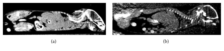
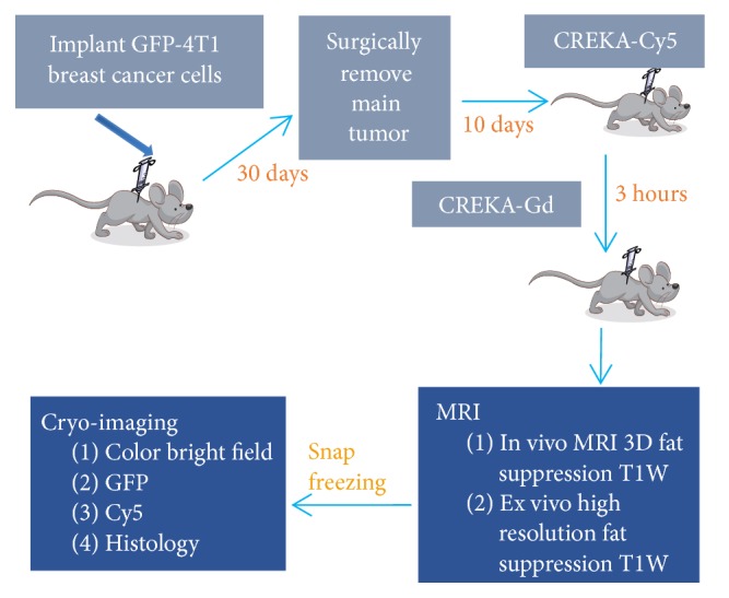
We created and evaluated a preclinical, multimodality imaging, and software platform to assess molecular imaging of small metastases. This included experimental methods (e.g., GFP-labeled tumor and high resolution multispectral cryo-imaging), nonrigid image registration, and interactive visualization of imaging agent targeting. We describe technological details earlier applied to GFP-labeled metastatic tumor targeting by molecular MR (CREKA-Gd) and red fluorescent (CREKA-Cy5) imaging agents. Optimized nonrigid cryo-MRI registration enabled nonambiguous association of MR signals to GFP tumors. Interactive visualization of out-of-RAM volumetric image data allowed one to zoom to a GFP-labeled micrometastasis, determine its anatomical location from color cryo-images, and establish the presence/absence of targeted CREKA-Gd and CREKA-Cy5. In a mouse with >160 GFP-labeled tumors, we determined that in the MR images every tumor in the lung >0.3 mm2 had visible signal and that some metastases as small as 0.1 mm2 were also visible. More tumors were visible in CREKA-Cy5 than in CREKA-Gd MRI. Tape transfer method and nonrigid registration allowed accurate (<11 μm error) registration of whole mouse histology to corresponding cryo-images. Histology showed inflammation and necrotic regions not labeled by imaging agents. This mouse-to-cells multiscale and multimodality platform should uniquely enable more informative and accurate studies of metastatic cancer imaging and therapy.
期刊介绍:
The International Journal of Biomedical Imaging is managed by a board of editors comprising internationally renowned active researchers. The journal is freely accessible online and also offered for purchase in print format. It employs a web-based review system to ensure swift turnaround times while maintaining high standards. In addition to regular issues, special issues are organized by guest editors. The subject areas covered include (but are not limited to):
Digital radiography and tomosynthesis
X-ray computed tomography (CT)
Magnetic resonance imaging (MRI)
Single photon emission computed tomography (SPECT)
Positron emission tomography (PET)
Ultrasound imaging
Diffuse optical tomography, coherence, fluorescence, bioluminescence tomography, impedance tomography
Neutron imaging for biomedical applications
Magnetic and optical spectroscopy, and optical biopsy
Optical, electron, scanning tunneling/atomic force microscopy
Small animal imaging
Functional, cellular, and molecular imaging
Imaging assays for screening and molecular analysis
Microarray image analysis and bioinformatics
Emerging biomedical imaging techniques
Imaging modality fusion
Biomedical imaging instrumentation
Biomedical image processing, pattern recognition, and analysis
Biomedical image visualization, compression, transmission, and storage
Imaging and modeling related to systems biology and systems biomedicine
Applied mathematics, applied physics, and chemistry related to biomedical imaging
Grid-enabling technology for biomedical imaging and informatics

 求助内容:
求助内容: 应助结果提醒方式:


