Tine Rosenberg, Charlotte Aaberg-Jessen, Stine Asferg Petterson, Bjarne Winther Kristensen
{"title":"长期缺氧条件下患者源性胶质母细胞瘤球形培养中干细胞标记物的异质表达。","authors":"Tine Rosenberg, Charlotte Aaberg-Jessen, Stine Asferg Petterson, Bjarne Winther Kristensen","doi":"10.2217/cns-2017-0034","DOIUrl":null,"url":null,"abstract":"<p><strong>Aim: </strong>To investigate the time profile of hypoxia and stem cell markers in glioblastoma spheroids of known molecular subtype.</p><p><strong>Materials & methods: </strong>Patient-derived glioblastoma spheroids were cultured up to 7 days in either 2% or 21% oxygen. Levels of proliferation (Ki-67), hypoxia (HIF-1α, CA9 and VEGF) and stem cell markers (CD133, nestin and musashi-1) were investigated by immunohistochemistry.</p><p><strong>Results: </strong>Hypoxia markers as well as CD133 and partially nestin increased in long-term hypoxia. The proliferation rate and spheroid size were highest in normoxia.</p><p><strong>Conclusion: </strong>We found differences in hypoxia and stem cell marker profiles between the patient-derived glioblastoma cultures. This heterogeneity should be taken into consideration in development of future therapeutic strategies.</p>","PeriodicalId":10469,"journal":{"name":"CNS Oncology","volume":"7 2","pages":"CNS15"},"PeriodicalIF":0.0000,"publicationDate":"2018-04-01","publicationTypes":"Journal Article","fieldsOfStudy":null,"isOpenAccess":false,"openAccessPdf":"https://sci-hub-pdf.com/10.2217/cns-2017-0034","citationCount":"6","resultStr":"{\"title\":\"Heterogenic expression of stem cell markers in patient-derived glioblastoma spheroid cultures exposed to long-term hypoxia.\",\"authors\":\"Tine Rosenberg, Charlotte Aaberg-Jessen, Stine Asferg Petterson, Bjarne Winther Kristensen\",\"doi\":\"10.2217/cns-2017-0034\",\"DOIUrl\":null,\"url\":null,\"abstract\":\"<p><strong>Aim: </strong>To investigate the time profile of hypoxia and stem cell markers in glioblastoma spheroids of known molecular subtype.</p><p><strong>Materials & methods: </strong>Patient-derived glioblastoma spheroids were cultured up to 7 days in either 2% or 21% oxygen. Levels of proliferation (Ki-67), hypoxia (HIF-1α, CA9 and VEGF) and stem cell markers (CD133, nestin and musashi-1) were investigated by immunohistochemistry.</p><p><strong>Results: </strong>Hypoxia markers as well as CD133 and partially nestin increased in long-term hypoxia. The proliferation rate and spheroid size were highest in normoxia.</p><p><strong>Conclusion: </strong>We found differences in hypoxia and stem cell marker profiles between the patient-derived glioblastoma cultures. This heterogeneity should be taken into consideration in development of future therapeutic strategies.</p>\",\"PeriodicalId\":10469,\"journal\":{\"name\":\"CNS Oncology\",\"volume\":\"7 2\",\"pages\":\"CNS15\"},\"PeriodicalIF\":0.0000,\"publicationDate\":\"2018-04-01\",\"publicationTypes\":\"Journal Article\",\"fieldsOfStudy\":null,\"isOpenAccess\":false,\"openAccessPdf\":\"https://sci-hub-pdf.com/10.2217/cns-2017-0034\",\"citationCount\":\"6\",\"resultStr\":null,\"platform\":\"Semanticscholar\",\"paperid\":null,\"PeriodicalName\":\"CNS Oncology\",\"FirstCategoryId\":\"1085\",\"ListUrlMain\":\"https://doi.org/10.2217/cns-2017-0034\",\"RegionNum\":0,\"RegionCategory\":null,\"ArticlePicture\":[],\"TitleCN\":null,\"AbstractTextCN\":null,\"PMCID\":null,\"EPubDate\":\"2018/4/30 0:00:00\",\"PubModel\":\"Epub\",\"JCR\":\"Q1\",\"JCRName\":\"Medicine\",\"Score\":null,\"Total\":0}","platform":"Semanticscholar","paperid":null,"PeriodicalName":"CNS Oncology","FirstCategoryId":"1085","ListUrlMain":"https://doi.org/10.2217/cns-2017-0034","RegionNum":0,"RegionCategory":null,"ArticlePicture":[],"TitleCN":null,"AbstractTextCN":null,"PMCID":null,"EPubDate":"2018/4/30 0:00:00","PubModel":"Epub","JCR":"Q1","JCRName":"Medicine","Score":null,"Total":0}
Heterogenic expression of stem cell markers in patient-derived glioblastoma spheroid cultures exposed to long-term hypoxia.
Aim: To investigate the time profile of hypoxia and stem cell markers in glioblastoma spheroids of known molecular subtype.
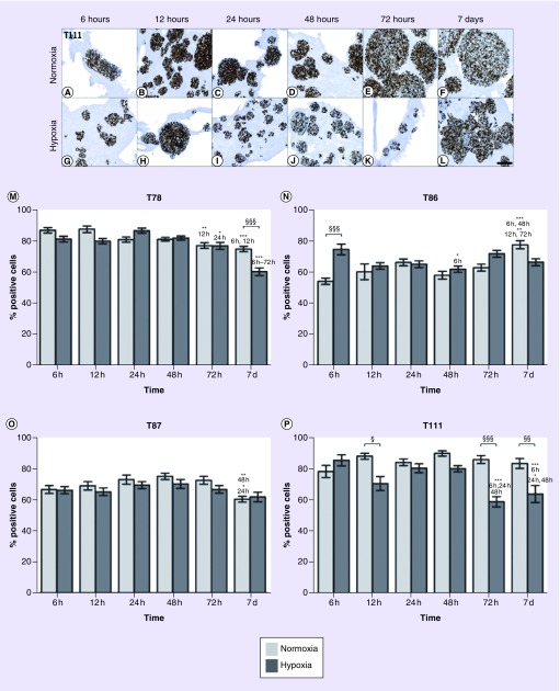
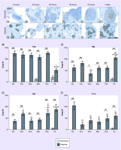
Materials & methods: Patient-derived glioblastoma spheroids were cultured up to 7 days in either 2% or 21% oxygen. Levels of proliferation (Ki-67), hypoxia (HIF-1α, CA9 and VEGF) and stem cell markers (CD133, nestin and musashi-1) were investigated by immunohistochemistry.
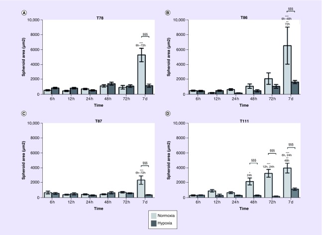
Results: Hypoxia markers as well as CD133 and partially nestin increased in long-term hypoxia. The proliferation rate and spheroid size were highest in normoxia.
Conclusion: We found differences in hypoxia and stem cell marker profiles between the patient-derived glioblastoma cultures. This heterogeneity should be taken into consideration in development of future therapeutic strategies.

 求助内容:
求助内容: 应助结果提醒方式:


