Renzo Guarnieri, Luca Testarelli, Luigi Stefanelli, Francesca De Angelis, Francesca Mencio, Giorgio Pompa, Stefano Di Carlo
{"title":"单独覆盖胶原膜或与猪源骨移植相关的拔牙槽骨愈合:组织学和组织形态学的比较分析。","authors":"Renzo Guarnieri, Luca Testarelli, Luigi Stefanelli, Francesca De Angelis, Francesca Mencio, Giorgio Pompa, Stefano Di Carlo","doi":"10.5037/jomr.2017.8404","DOIUrl":null,"url":null,"abstract":"<p><strong>Objectives: </strong>The present paper reports data of a randomized study aimed to analyse and compare the histologic and histomorphometric aspects of bone healing in extraction sites covered with collagen membrane alone or associated with porcine-derived bone graft.</p><p><strong>Material and methods: </strong>Thirty patients, with single extraction sockets without severe bone wall defects in the premolar/molar region, were included. Ten extraction sockets were grafted with porcine-derived bone and covered with collagen membrane (group 1), 10 sites were covered with collagen membrane alone (group 2), and 10 sites healed spontaneously (group 3). After 4 months of healing, 26 (8 in group 1, 9 in group 2, and 9 in group 3) bone core specimens were harvested for histologic evaluation, then dental implants were placed.</p><p><strong>Results: </strong>Sites in the group 1 and in the group 2 showed similar histologic and histomorphometric results without significantly differences in the percentage of vital bone (57.43% [SD 4.8] vs. 60.01% [SD 3.2]), and non-mineralized connective tissue 22.99% (SD 5.3) vs. 18.53% (SD 6.2). In group 1 a 16.57% (SD 3.8) of residual material was found.</p><p><strong>Conclusions: </strong>Results showed that the use of collagen membrane alone or associated to porcine-derived bone improves the healing bone process compared to that of extraction sites spontaneously healed. Moreover, histomorphometric data related to bone quality, indicated that extraction sites without severe walls defects and with a vestibular bone thickness > 1.5 mm, treated with a low resorbtion rate collagen membrane alone, do not need more than 4 months for dental implant insertion.</p>","PeriodicalId":230885,"journal":{"name":"Journal of Oral & Maxillofacial Research","volume":"8 4","pages":"e4"},"PeriodicalIF":0.0000,"publicationDate":"2017-12-31","publicationTypes":"Journal Article","fieldsOfStudy":null,"isOpenAccess":false,"openAccessPdf":"https://sci-hub-pdf.com/10.5037/jomr.2017.8404","citationCount":"15","resultStr":"{\"title\":\"Bone Healing in Extraction Sockets Covered With Collagen Membrane Alone or Associated With Porcine-Derived Bone Graft: a Comparative Histological and Histomorphometric Analysis.\",\"authors\":\"Renzo Guarnieri, Luca Testarelli, Luigi Stefanelli, Francesca De Angelis, Francesca Mencio, Giorgio Pompa, Stefano Di Carlo\",\"doi\":\"10.5037/jomr.2017.8404\",\"DOIUrl\":null,\"url\":null,\"abstract\":\"<p><strong>Objectives: </strong>The present paper reports data of a randomized study aimed to analyse and compare the histologic and histomorphometric aspects of bone healing in extraction sites covered with collagen membrane alone or associated with porcine-derived bone graft.</p><p><strong>Material and methods: </strong>Thirty patients, with single extraction sockets without severe bone wall defects in the premolar/molar region, were included. Ten extraction sockets were grafted with porcine-derived bone and covered with collagen membrane (group 1), 10 sites were covered with collagen membrane alone (group 2), and 10 sites healed spontaneously (group 3). After 4 months of healing, 26 (8 in group 1, 9 in group 2, and 9 in group 3) bone core specimens were harvested for histologic evaluation, then dental implants were placed.</p><p><strong>Results: </strong>Sites in the group 1 and in the group 2 showed similar histologic and histomorphometric results without significantly differences in the percentage of vital bone (57.43% [SD 4.8] vs. 60.01% [SD 3.2]), and non-mineralized connective tissue 22.99% (SD 5.3) vs. 18.53% (SD 6.2). In group 1 a 16.57% (SD 3.8) of residual material was found.</p><p><strong>Conclusions: </strong>Results showed that the use of collagen membrane alone or associated to porcine-derived bone improves the healing bone process compared to that of extraction sites spontaneously healed. Moreover, histomorphometric data related to bone quality, indicated that extraction sites without severe walls defects and with a vestibular bone thickness > 1.5 mm, treated with a low resorbtion rate collagen membrane alone, do not need more than 4 months for dental implant insertion.</p>\",\"PeriodicalId\":230885,\"journal\":{\"name\":\"Journal of Oral & Maxillofacial Research\",\"volume\":\"8 4\",\"pages\":\"e4\"},\"PeriodicalIF\":0.0000,\"publicationDate\":\"2017-12-31\",\"publicationTypes\":\"Journal Article\",\"fieldsOfStudy\":null,\"isOpenAccess\":false,\"openAccessPdf\":\"https://sci-hub-pdf.com/10.5037/jomr.2017.8404\",\"citationCount\":\"15\",\"resultStr\":null,\"platform\":\"Semanticscholar\",\"paperid\":null,\"PeriodicalName\":\"Journal of Oral & Maxillofacial Research\",\"FirstCategoryId\":\"1085\",\"ListUrlMain\":\"https://doi.org/10.5037/jomr.2017.8404\",\"RegionNum\":0,\"RegionCategory\":null,\"ArticlePicture\":[],\"TitleCN\":null,\"AbstractTextCN\":null,\"PMCID\":null,\"EPubDate\":\"2017/10/1 0:00:00\",\"PubModel\":\"eCollection\",\"JCR\":\"\",\"JCRName\":\"\",\"Score\":null,\"Total\":0}","platform":"Semanticscholar","paperid":null,"PeriodicalName":"Journal of Oral & Maxillofacial Research","FirstCategoryId":"1085","ListUrlMain":"https://doi.org/10.5037/jomr.2017.8404","RegionNum":0,"RegionCategory":null,"ArticlePicture":[],"TitleCN":null,"AbstractTextCN":null,"PMCID":null,"EPubDate":"2017/10/1 0:00:00","PubModel":"eCollection","JCR":"","JCRName":"","Score":null,"Total":0}
引用次数: 15
摘要
目的:本文报告了一项随机研究的数据,旨在分析和比较单独覆盖胶原膜或与猪源性骨移植物相关的提取部位骨愈合的组织学和组织形态学方面。材料与方法:选取前磨牙/磨牙区无严重骨壁缺损的单牙槽位患者30例。10个拔牙槽骨采用猪源性骨并覆盖胶原膜(1组),10个部位单独覆盖胶原膜(2组),10个部位自行愈合(3组)。愈合4个月后,取26个(1组8个,2组9个,3组9个)骨核标本进行组织学评估,放置种植体。结果:1组和2组的组织学和组织形态测量结果相似,但在活骨百分比(57.43% [SD 4.8] vs. 60.01% [SD 3.2])和非矿化结缔组织百分比(22.99% (SD 5.3) vs. 18.53% (SD 6.2))方面无显著差异。1组残留物质16.57% (SD 3.8)。结论:结果表明,与提取部位自发愈合相比,单独使用胶原膜或与猪源性骨联合使用可改善骨愈合过程。此外,与骨质量相关的组织形态学数据表明,拔牙部位没有严重的壁缺损,前庭骨厚度> 1.5 mm,单独使用低吸收率胶原膜处理,种植牙插入时间不超过4个月。
Bone Healing in Extraction Sockets Covered With Collagen Membrane Alone or Associated With Porcine-Derived Bone Graft: a Comparative Histological and Histomorphometric Analysis.
Objectives: The present paper reports data of a randomized study aimed to analyse and compare the histologic and histomorphometric aspects of bone healing in extraction sites covered with collagen membrane alone or associated with porcine-derived bone graft.
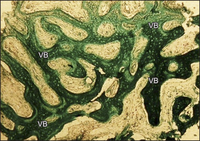
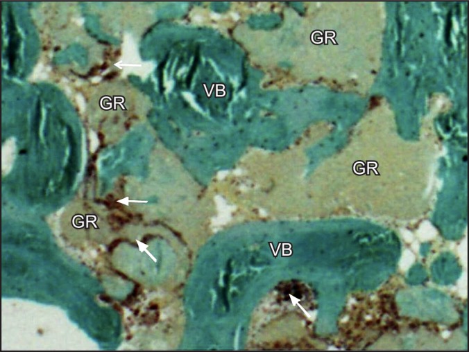
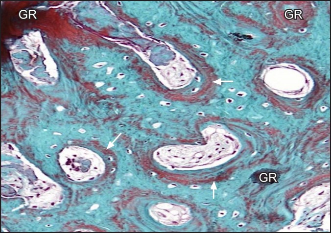
Material and methods: Thirty patients, with single extraction sockets without severe bone wall defects in the premolar/molar region, were included. Ten extraction sockets were grafted with porcine-derived bone and covered with collagen membrane (group 1), 10 sites were covered with collagen membrane alone (group 2), and 10 sites healed spontaneously (group 3). After 4 months of healing, 26 (8 in group 1, 9 in group 2, and 9 in group 3) bone core specimens were harvested for histologic evaluation, then dental implants were placed.
Results: Sites in the group 1 and in the group 2 showed similar histologic and histomorphometric results without significantly differences in the percentage of vital bone (57.43% [SD 4.8] vs. 60.01% [SD 3.2]), and non-mineralized connective tissue 22.99% (SD 5.3) vs. 18.53% (SD 6.2). In group 1 a 16.57% (SD 3.8) of residual material was found.
Conclusions: Results showed that the use of collagen membrane alone or associated to porcine-derived bone improves the healing bone process compared to that of extraction sites spontaneously healed. Moreover, histomorphometric data related to bone quality, indicated that extraction sites without severe walls defects and with a vestibular bone thickness > 1.5 mm, treated with a low resorbtion rate collagen membrane alone, do not need more than 4 months for dental implant insertion.

 求助内容:
求助内容: 应助结果提醒方式:


