Heba Radi AttaAllah, Ismail Ahmed Nagib Omar, Ahmed Shawkat Abdelhalim
{"title":"光谱域OCT对高度近视眼后段的评价。","authors":"Heba Radi AttaAllah, Ismail Ahmed Nagib Omar, Ahmed Shawkat Abdelhalim","doi":"10.2174/1874364101711010334","DOIUrl":null,"url":null,"abstract":"<p><strong>Purpose: </strong>Spectral Domain Optical Coherence Tomography (SD-OCT) was used to evaluate retinal and vitreo-retinal changes that occur in highly myopic patients.</p><p><strong>Methods: </strong>This prospective study included 472 eyes of 472 patients suffering from high myopia (> -6.00 D), between May 2012 and December 2015. All patients were examined, using Cirrus HD OCT (Zeiss Cirrus TM HD-OCT model 4000), to detect any retinal or vitreo-retinal interface abnormalities.All obtained data was analyzed using Statistical Package for the Social Sciences software version 17 (SPSS Inc, Chicago, IL, USA) and the paired two-sided t-test. Bivariate correlations were performed between different parameters using the Spearman correlation coefficient (r).</p><p><strong>Results: </strong>Mean spherical equivalent (MSE) was -13.11± 4.35D. Mean axial length (AL) was 28.5±1.62 mm. Posterior vitreous detachment (PVD) was the most frequent OCT finding; representing 33.4% of the cases, 13.7% of them were associated with macular traction. A statistically significant positive correlation was found between AL and MTM, full thickness macular hole, PVD with traction, and dome shaped macula (r = 0.49 and P = 0.001, r = 0.422 and P = 0.0001, r = 0.25 and P = 0.03, r=0.475, P=0.001 respectively).</p><p><strong>Conclusion: </strong>OCT is a valuable tool in detecting retinal and vitreo-retinal interface abnormalities in highly myopic eyes, and it can be used for follow up of those patients to avoid advanced retinal damage.</p>","PeriodicalId":512318,"journal":{"name":"The Open Ophthalmology Journal","volume":"11 ","pages":"334-345"},"PeriodicalIF":0.0000,"publicationDate":"2017-11-22","publicationTypes":"Journal Article","fieldsOfStudy":null,"isOpenAccess":false,"openAccessPdf":"https://www.ncbi.nlm.nih.gov/pmc/articles/PMC5725562/pdf/","citationCount":"10","resultStr":"{\"title\":\"Assessment of Posterior Segment Using Spectral Domain OCT in Highly Myopic Eyes.\",\"authors\":\"Heba Radi AttaAllah, Ismail Ahmed Nagib Omar, Ahmed Shawkat Abdelhalim\",\"doi\":\"10.2174/1874364101711010334\",\"DOIUrl\":null,\"url\":null,\"abstract\":\"<p><strong>Purpose: </strong>Spectral Domain Optical Coherence Tomography (SD-OCT) was used to evaluate retinal and vitreo-retinal changes that occur in highly myopic patients.</p><p><strong>Methods: </strong>This prospective study included 472 eyes of 472 patients suffering from high myopia (> -6.00 D), between May 2012 and December 2015. All patients were examined, using Cirrus HD OCT (Zeiss Cirrus TM HD-OCT model 4000), to detect any retinal or vitreo-retinal interface abnormalities.All obtained data was analyzed using Statistical Package for the Social Sciences software version 17 (SPSS Inc, Chicago, IL, USA) and the paired two-sided t-test. Bivariate correlations were performed between different parameters using the Spearman correlation coefficient (r).</p><p><strong>Results: </strong>Mean spherical equivalent (MSE) was -13.11± 4.35D. Mean axial length (AL) was 28.5±1.62 mm. Posterior vitreous detachment (PVD) was the most frequent OCT finding; representing 33.4% of the cases, 13.7% of them were associated with macular traction. A statistically significant positive correlation was found between AL and MTM, full thickness macular hole, PVD with traction, and dome shaped macula (r = 0.49 and P = 0.001, r = 0.422 and P = 0.0001, r = 0.25 and P = 0.03, r=0.475, P=0.001 respectively).</p><p><strong>Conclusion: </strong>OCT is a valuable tool in detecting retinal and vitreo-retinal interface abnormalities in highly myopic eyes, and it can be used for follow up of those patients to avoid advanced retinal damage.</p>\",\"PeriodicalId\":512318,\"journal\":{\"name\":\"The Open Ophthalmology Journal\",\"volume\":\"11 \",\"pages\":\"334-345\"},\"PeriodicalIF\":0.0000,\"publicationDate\":\"2017-11-22\",\"publicationTypes\":\"Journal Article\",\"fieldsOfStudy\":null,\"isOpenAccess\":false,\"openAccessPdf\":\"https://www.ncbi.nlm.nih.gov/pmc/articles/PMC5725562/pdf/\",\"citationCount\":\"10\",\"resultStr\":null,\"platform\":\"Semanticscholar\",\"paperid\":null,\"PeriodicalName\":\"The Open Ophthalmology Journal\",\"FirstCategoryId\":\"1085\",\"ListUrlMain\":\"https://doi.org/10.2174/1874364101711010334\",\"RegionNum\":0,\"RegionCategory\":null,\"ArticlePicture\":[],\"TitleCN\":null,\"AbstractTextCN\":null,\"PMCID\":null,\"EPubDate\":\"2017/1/1 0:00:00\",\"PubModel\":\"eCollection\",\"JCR\":\"\",\"JCRName\":\"\",\"Score\":null,\"Total\":0}","platform":"Semanticscholar","paperid":null,"PeriodicalName":"The Open Ophthalmology Journal","FirstCategoryId":"1085","ListUrlMain":"https://doi.org/10.2174/1874364101711010334","RegionNum":0,"RegionCategory":null,"ArticlePicture":[],"TitleCN":null,"AbstractTextCN":null,"PMCID":null,"EPubDate":"2017/1/1 0:00:00","PubModel":"eCollection","JCR":"","JCRName":"","Score":null,"Total":0}
Assessment of Posterior Segment Using Spectral Domain OCT in Highly Myopic Eyes.
Purpose: Spectral Domain Optical Coherence Tomography (SD-OCT) was used to evaluate retinal and vitreo-retinal changes that occur in highly myopic patients.
Methods: This prospective study included 472 eyes of 472 patients suffering from high myopia (> -6.00 D), between May 2012 and December 2015. All patients were examined, using Cirrus HD OCT (Zeiss Cirrus TM HD-OCT model 4000), to detect any retinal or vitreo-retinal interface abnormalities.All obtained data was analyzed using Statistical Package for the Social Sciences software version 17 (SPSS Inc, Chicago, IL, USA) and the paired two-sided t-test. Bivariate correlations were performed between different parameters using the Spearman correlation coefficient (r).
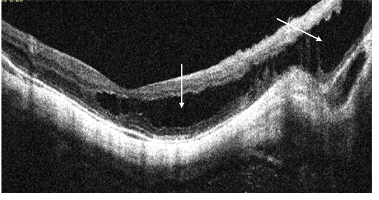
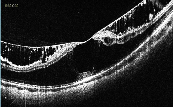
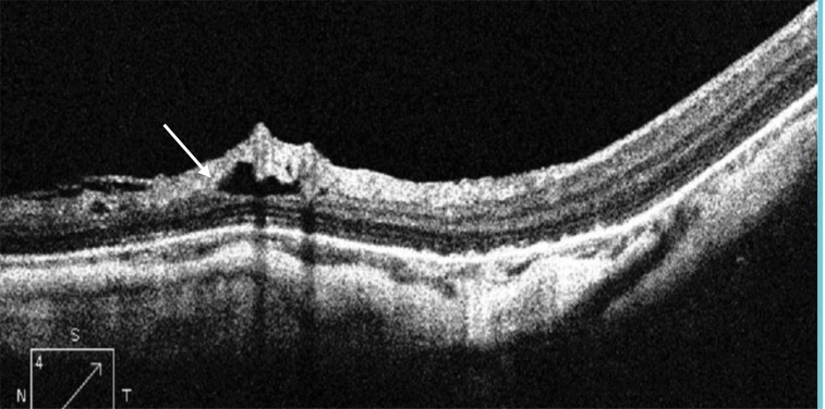
Results: Mean spherical equivalent (MSE) was -13.11± 4.35D. Mean axial length (AL) was 28.5±1.62 mm. Posterior vitreous detachment (PVD) was the most frequent OCT finding; representing 33.4% of the cases, 13.7% of them were associated with macular traction. A statistically significant positive correlation was found between AL and MTM, full thickness macular hole, PVD with traction, and dome shaped macula (r = 0.49 and P = 0.001, r = 0.422 and P = 0.0001, r = 0.25 and P = 0.03, r=0.475, P=0.001 respectively).
Conclusion: OCT is a valuable tool in detecting retinal and vitreo-retinal interface abnormalities in highly myopic eyes, and it can be used for follow up of those patients to avoid advanced retinal damage.

 求助内容:
求助内容: 应助结果提醒方式:


