Renzo Guarnieri, Luigi Stefanelli, Francesca De Angelis, Francesca Mencio, Giorgio Pompa, Stefano Di Carlo
{"title":"单独使用或与人造茯苓骨结合使用茯苓胶原膜保存拔牙窝。随机对照研究的临床结果。","authors":"Renzo Guarnieri, Luigi Stefanelli, Francesca De Angelis, Francesca Mencio, Giorgio Pompa, Stefano Di Carlo","doi":"10.5037/jomr.2017.8305","DOIUrl":null,"url":null,"abstract":"<p><strong>Objectives: </strong>The aim of present randomized controlled clinical trial was to clinically evaluate hard tissue changes after extraction socket preservation procedures compared to natural spontaneous healing.</p><p><strong>Material and methods: </strong>Thirty patients were enrolled in the present study and underwent single-tooth extraction in the premolar/molar areas. Ten sites were grafted with porcine-derived bone covered by collagen membrane, 10 covered by porcine-derived collagen membrane alone, and 10 underwent natural spontaneous healing. Vertical and horizontal bone changes after 3-month were evaluated at implant placement.</p><p><strong>Results: </strong>The vertical and horizontal bone changes at the extraction sockets treated with collagen membrane alone (vertical: -0.55 [SD 0.11] mm, and horizontal: -1.21 [SD 0.69] mm) and collagen membrane plus porcine-derived bone (vertical: -0.37 [SD 0.7] mm, and horizontal: -0.91 [SD 0.53] mm) were found significantly lower (P < 0.001), when compared to non-grafted sockets (vertical: -2.09 [SD 0.19] mm, and horizontal: -3.96 [SD 0.87] mm).In type 1 extraction sockets, in premolar sites, and in presence of vestibular bone thicknesses ≥ 1.5 mm, the use of collagen membrane alone revealed similar outcomes to those with additional graft material.</p><p><strong>Conclusions: </strong>At the re-entry surgery, extraction sockets grafted with porcine-derived bone and covered by collagen membrane, and extraction sockets covered by porcine-derived collagen membrane alone, showed significantly lower vertical and horizontal bone changes, compared to extraction sockets sites underwent natural spontaneous healing. However, a complete prevention of remodelling is not achievable, irrespective of the technique used.</p>","PeriodicalId":53254,"journal":{"name":"eJournal of Oral Maxillofacial Research","volume":"8 3","pages":"e5"},"PeriodicalIF":1.0000,"publicationDate":"2017-09-30","publicationTypes":"Journal Article","fieldsOfStudy":null,"isOpenAccess":false,"openAccessPdf":"https://ftp.ncbi.nlm.nih.gov/pub/pmc/oa_pdf/c2/4c/jomr-08-e5.PMC5676315.pdf","citationCount":"0","resultStr":"{\"title\":\"Extraction Socket Preservation Using Porcine-Derived Collagen Membrane Alone or Associated with Porcine-Derived Bone. Clinical Results of Randomized Controlled Study.\",\"authors\":\"Renzo Guarnieri, Luigi Stefanelli, Francesca De Angelis, Francesca Mencio, Giorgio Pompa, Stefano Di Carlo\",\"doi\":\"10.5037/jomr.2017.8305\",\"DOIUrl\":null,\"url\":null,\"abstract\":\"<p><strong>Objectives: </strong>The aim of present randomized controlled clinical trial was to clinically evaluate hard tissue changes after extraction socket preservation procedures compared to natural spontaneous healing.</p><p><strong>Material and methods: </strong>Thirty patients were enrolled in the present study and underwent single-tooth extraction in the premolar/molar areas. Ten sites were grafted with porcine-derived bone covered by collagen membrane, 10 covered by porcine-derived collagen membrane alone, and 10 underwent natural spontaneous healing. Vertical and horizontal bone changes after 3-month were evaluated at implant placement.</p><p><strong>Results: </strong>The vertical and horizontal bone changes at the extraction sockets treated with collagen membrane alone (vertical: -0.55 [SD 0.11] mm, and horizontal: -1.21 [SD 0.69] mm) and collagen membrane plus porcine-derived bone (vertical: -0.37 [SD 0.7] mm, and horizontal: -0.91 [SD 0.53] mm) were found significantly lower (P < 0.001), when compared to non-grafted sockets (vertical: -2.09 [SD 0.19] mm, and horizontal: -3.96 [SD 0.87] mm).In type 1 extraction sockets, in premolar sites, and in presence of vestibular bone thicknesses ≥ 1.5 mm, the use of collagen membrane alone revealed similar outcomes to those with additional graft material.</p><p><strong>Conclusions: </strong>At the re-entry surgery, extraction sockets grafted with porcine-derived bone and covered by collagen membrane, and extraction sockets covered by porcine-derived collagen membrane alone, showed significantly lower vertical and horizontal bone changes, compared to extraction sockets sites underwent natural spontaneous healing. However, a complete prevention of remodelling is not achievable, irrespective of the technique used.</p>\",\"PeriodicalId\":53254,\"journal\":{\"name\":\"eJournal of Oral Maxillofacial Research\",\"volume\":\"8 3\",\"pages\":\"e5\"},\"PeriodicalIF\":1.0000,\"publicationDate\":\"2017-09-30\",\"publicationTypes\":\"Journal Article\",\"fieldsOfStudy\":null,\"isOpenAccess\":false,\"openAccessPdf\":\"https://ftp.ncbi.nlm.nih.gov/pub/pmc/oa_pdf/c2/4c/jomr-08-e5.PMC5676315.pdf\",\"citationCount\":\"0\",\"resultStr\":null,\"platform\":\"Semanticscholar\",\"paperid\":null,\"PeriodicalName\":\"eJournal of Oral Maxillofacial Research\",\"FirstCategoryId\":\"1085\",\"ListUrlMain\":\"https://doi.org/10.5037/jomr.2017.8305\",\"RegionNum\":0,\"RegionCategory\":null,\"ArticlePicture\":[],\"TitleCN\":null,\"AbstractTextCN\":null,\"PMCID\":null,\"EPubDate\":\"2017/7/1 0:00:00\",\"PubModel\":\"eCollection\",\"JCR\":\"Q3\",\"JCRName\":\"DENTISTRY, ORAL SURGERY & MEDICINE\",\"Score\":null,\"Total\":0}","platform":"Semanticscholar","paperid":null,"PeriodicalName":"eJournal of Oral Maxillofacial Research","FirstCategoryId":"1085","ListUrlMain":"https://doi.org/10.5037/jomr.2017.8305","RegionNum":0,"RegionCategory":null,"ArticlePicture":[],"TitleCN":null,"AbstractTextCN":null,"PMCID":null,"EPubDate":"2017/7/1 0:00:00","PubModel":"eCollection","JCR":"Q3","JCRName":"DENTISTRY, ORAL SURGERY & MEDICINE","Score":null,"Total":0}
Extraction Socket Preservation Using Porcine-Derived Collagen Membrane Alone or Associated with Porcine-Derived Bone. Clinical Results of Randomized Controlled Study.
Objectives: The aim of present randomized controlled clinical trial was to clinically evaluate hard tissue changes after extraction socket preservation procedures compared to natural spontaneous healing.
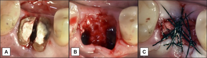
Material and methods: Thirty patients were enrolled in the present study and underwent single-tooth extraction in the premolar/molar areas. Ten sites were grafted with porcine-derived bone covered by collagen membrane, 10 covered by porcine-derived collagen membrane alone, and 10 underwent natural spontaneous healing. Vertical and horizontal bone changes after 3-month were evaluated at implant placement.
Results: The vertical and horizontal bone changes at the extraction sockets treated with collagen membrane alone (vertical: -0.55 [SD 0.11] mm, and horizontal: -1.21 [SD 0.69] mm) and collagen membrane plus porcine-derived bone (vertical: -0.37 [SD 0.7] mm, and horizontal: -0.91 [SD 0.53] mm) were found significantly lower (P < 0.001), when compared to non-grafted sockets (vertical: -2.09 [SD 0.19] mm, and horizontal: -3.96 [SD 0.87] mm).In type 1 extraction sockets, in premolar sites, and in presence of vestibular bone thicknesses ≥ 1.5 mm, the use of collagen membrane alone revealed similar outcomes to those with additional graft material.
Conclusions: At the re-entry surgery, extraction sockets grafted with porcine-derived bone and covered by collagen membrane, and extraction sockets covered by porcine-derived collagen membrane alone, showed significantly lower vertical and horizontal bone changes, compared to extraction sockets sites underwent natural spontaneous healing. However, a complete prevention of remodelling is not achievable, irrespective of the technique used.

 求助内容:
求助内容: 应助结果提醒方式:


