{"title":"二十二碳六烯酸抑制葡萄糖变异性诱导的血管平滑肌细胞增殖。","authors":"Kaliyaperumal Rani, Nway Y Aung","doi":"10.2174/1874091X01711010056","DOIUrl":null,"url":null,"abstract":"<p><strong>Background: </strong>Vascular Smooth Muscle cells (VSMC) enact crucial roles in early vasculogenesis and sustenance of vascular integrity. However, aberrant proliferation of VSMC followed by migration into the blood vessel wall leads to the formation of vascular lesions accounting for the degeneration and remodelling of vascular basement membrane. In diabetes, hyperglycaemia accelerates VSMC proliferation and contributes to the initiation and progression of atherosclerotic lesions. Recently, acute glucose fluctuations have been implicated in the abnormal VSMC proliferation and complications of diabetic atherosclerosis. Docosahexaenoic acid (DHA), a ω-3 polyunsaturated fatty acid (PUFA) has been shown to inhibit proliferation of several cell types implicating several different mechanisms. In the present study, we have investigated the effects of DHA on VSMC proliferation induced by stable and intermittent high glucose levels.</p><p><strong>Method: </strong>Confluent cultures of rat aortic VSMCs were treated with DHA for 24 hrs and then exposed to stable high glucose (25 mmol/L, SHG) or intermittent high glucose (5 mmol/L and 25 mmol/L alternating every 12 hrs, IHG) for 72 hrs. Cell proliferation was examined by the MTT viability assay, while apoptosis process was evaluated by the Hoechst staining, flow cytometry and caspase-3 activity assays.</p><p><strong>Results: </strong>Our data demonstrated that the hyper proliferation induced by stable and intermittent high glucose levels was significantly inhibited by the DHA pre-treatment. DHA significantly increased caspase-3 activity, resulting in enhanced DNA fragmentation and apoptosis.</p><p><strong>Conclusion: </strong>Our results suggest that DHA reduced the high glucose-induced proliferation of VSMC and induced cell apoptosis.</p>","PeriodicalId":515405,"journal":{"name":"The Open Biochemistry Journal","volume":"11 ","pages":"56-65"},"PeriodicalIF":0.0000,"publicationDate":"2017-06-30","publicationTypes":"Journal Article","fieldsOfStudy":null,"isOpenAccess":false,"openAccessPdf":"https://www.ncbi.nlm.nih.gov/pmc/articles/PMC5543665/pdf/","citationCount":"11","resultStr":"{\"title\":\"Docosahexaenoic Acid Inhibits Vascular Smooth Muscle Cell Proliferation Induced by Glucose Variability.\",\"authors\":\"Kaliyaperumal Rani, Nway Y Aung\",\"doi\":\"10.2174/1874091X01711010056\",\"DOIUrl\":null,\"url\":null,\"abstract\":\"<p><strong>Background: </strong>Vascular Smooth Muscle cells (VSMC) enact crucial roles in early vasculogenesis and sustenance of vascular integrity. However, aberrant proliferation of VSMC followed by migration into the blood vessel wall leads to the formation of vascular lesions accounting for the degeneration and remodelling of vascular basement membrane. In diabetes, hyperglycaemia accelerates VSMC proliferation and contributes to the initiation and progression of atherosclerotic lesions. Recently, acute glucose fluctuations have been implicated in the abnormal VSMC proliferation and complications of diabetic atherosclerosis. Docosahexaenoic acid (DHA), a ω-3 polyunsaturated fatty acid (PUFA) has been shown to inhibit proliferation of several cell types implicating several different mechanisms. In the present study, we have investigated the effects of DHA on VSMC proliferation induced by stable and intermittent high glucose levels.</p><p><strong>Method: </strong>Confluent cultures of rat aortic VSMCs were treated with DHA for 24 hrs and then exposed to stable high glucose (25 mmol/L, SHG) or intermittent high glucose (5 mmol/L and 25 mmol/L alternating every 12 hrs, IHG) for 72 hrs. Cell proliferation was examined by the MTT viability assay, while apoptosis process was evaluated by the Hoechst staining, flow cytometry and caspase-3 activity assays.</p><p><strong>Results: </strong>Our data demonstrated that the hyper proliferation induced by stable and intermittent high glucose levels was significantly inhibited by the DHA pre-treatment. DHA significantly increased caspase-3 activity, resulting in enhanced DNA fragmentation and apoptosis.</p><p><strong>Conclusion: </strong>Our results suggest that DHA reduced the high glucose-induced proliferation of VSMC and induced cell apoptosis.</p>\",\"PeriodicalId\":515405,\"journal\":{\"name\":\"The Open Biochemistry Journal\",\"volume\":\"11 \",\"pages\":\"56-65\"},\"PeriodicalIF\":0.0000,\"publicationDate\":\"2017-06-30\",\"publicationTypes\":\"Journal Article\",\"fieldsOfStudy\":null,\"isOpenAccess\":false,\"openAccessPdf\":\"https://www.ncbi.nlm.nih.gov/pmc/articles/PMC5543665/pdf/\",\"citationCount\":\"11\",\"resultStr\":null,\"platform\":\"Semanticscholar\",\"paperid\":null,\"PeriodicalName\":\"The Open Biochemistry Journal\",\"FirstCategoryId\":\"1085\",\"ListUrlMain\":\"https://doi.org/10.2174/1874091X01711010056\",\"RegionNum\":0,\"RegionCategory\":null,\"ArticlePicture\":[],\"TitleCN\":null,\"AbstractTextCN\":null,\"PMCID\":null,\"EPubDate\":\"2017/1/1 0:00:00\",\"PubModel\":\"eCollection\",\"JCR\":\"\",\"JCRName\":\"\",\"Score\":null,\"Total\":0}","platform":"Semanticscholar","paperid":null,"PeriodicalName":"The Open Biochemistry Journal","FirstCategoryId":"1085","ListUrlMain":"https://doi.org/10.2174/1874091X01711010056","RegionNum":0,"RegionCategory":null,"ArticlePicture":[],"TitleCN":null,"AbstractTextCN":null,"PMCID":null,"EPubDate":"2017/1/1 0:00:00","PubModel":"eCollection","JCR":"","JCRName":"","Score":null,"Total":0}
Background: Vascular Smooth Muscle cells (VSMC) enact crucial roles in early vasculogenesis and sustenance of vascular integrity. However, aberrant proliferation of VSMC followed by migration into the blood vessel wall leads to the formation of vascular lesions accounting for the degeneration and remodelling of vascular basement membrane. In diabetes, hyperglycaemia accelerates VSMC proliferation and contributes to the initiation and progression of atherosclerotic lesions. Recently, acute glucose fluctuations have been implicated in the abnormal VSMC proliferation and complications of diabetic atherosclerosis. Docosahexaenoic acid (DHA), a ω-3 polyunsaturated fatty acid (PUFA) has been shown to inhibit proliferation of several cell types implicating several different mechanisms. In the present study, we have investigated the effects of DHA on VSMC proliferation induced by stable and intermittent high glucose levels.
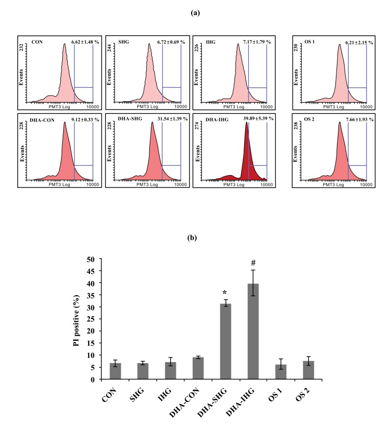
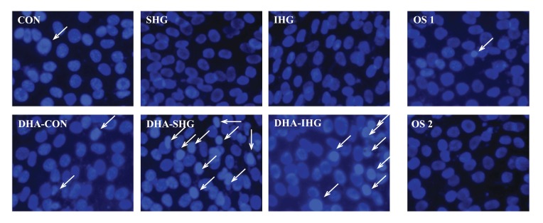
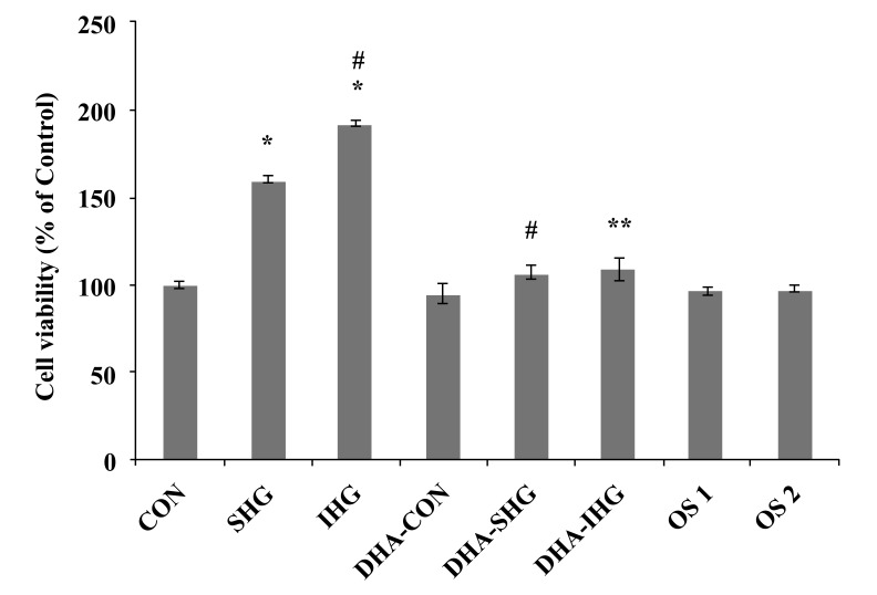
Method: Confluent cultures of rat aortic VSMCs were treated with DHA for 24 hrs and then exposed to stable high glucose (25 mmol/L, SHG) or intermittent high glucose (5 mmol/L and 25 mmol/L alternating every 12 hrs, IHG) for 72 hrs. Cell proliferation was examined by the MTT viability assay, while apoptosis process was evaluated by the Hoechst staining, flow cytometry and caspase-3 activity assays.
Results: Our data demonstrated that the hyper proliferation induced by stable and intermittent high glucose levels was significantly inhibited by the DHA pre-treatment. DHA significantly increased caspase-3 activity, resulting in enhanced DNA fragmentation and apoptosis.
Conclusion: Our results suggest that DHA reduced the high glucose-induced proliferation of VSMC and induced cell apoptosis.

 求助内容:
求助内容: 应助结果提醒方式:


