{"title":"系统性脑膜炎球菌病患者死后组织样本中脑膜炎奈瑟菌DNA的可追溯性和分布","authors":"Berit Sletbakk Brusletto, Bernt Christian Hellerud, Else Marit Løberg, Ingeborg Løstegaard Goverud, Åshild Vege, Jens Petter Berg, Petter Brandtzaeg, Reidun Øvstebø","doi":"10.1186/s12907-017-0049-9","DOIUrl":null,"url":null,"abstract":"<p><strong>Background: </strong>The pathophysiology and outcome of meningococcal septic shock is closely associated with the plasma level of <i>N. meningitidis</i> lipopolysaccharides (LPS, endotoxin) and the circulating level of meningococcal DNA. The aim of the present study was to quantify the number of <i>N. meningitidis</i> in different formalin-fixed, paraffin-embedded (FFPE) tissue samples and fresh frozen (FF) tissue samples from patients with systemic meningococcal disease (SMD), to explore the distribution of <i>N. meningitidis</i> in the body.</p><p><strong>Methods: </strong>DNA in FFPE and FF tissue samples from heart, lungs, liver, kidneys, spleen and brain from patients with meningococcal shock and controls (lethal pneumococcal infection) stored at variable times, were isolated. The bacterial load of <i>N. meningitidis</i> DNA was analyzed using quantitative real-time PCR (qPCR) and primers for the capsule transport A (ctrA) gene (1 copy per <i>N. meningitidis</i> DNA). The human beta-hemoglobin (HBB) gene was quantified to evaluate effect of the storage times (2-28 years) and storage method in archived tissue.</p><p><strong>Results: </strong><i>N. meningitidis</i> DNA was detected in FFPE and FF tissue samples from heart, lung, liver, kidney, and spleen in all patients with severe shock. In FFPE brain, <i>N. meningitidis</i> DNA was only detected in the patient with the highest concentration of LPS in the blood at admission to hospital. The highest levels of <i>N. meningitidis</i> DNA were found in heart tissue (median value 3.6 × 10<sup>7</sup> copies <i>N. meningitidis</i> DNA/μg human DNA) and lung tissue (median value 3.1 × 10<sup>7</sup> copies <i>N. meningitidis</i> DNA/μg human DNA) in all five patients. <i>N. meningitidis</i> DNA was not detectable in any of the tissue samples from two patients with clinical meningitis and the controls (pneumococcal infection). The quantity of HBB declined over time in FFPE tissue stored at room temperature, suggesting degradation of DNA.</p><p><strong>Conclusions: </strong>High levels of <i>N. meningitidis</i> DNA were detected in the different tissue samples from meningococcal shock patients, particularly in the heart and lungs suggesting seeding and major proliferation of meningococci in these organs during the development of shock, probably contributing to the multiple organ failure. The age of archived tissue samples appear to have an impact on the amount of quantifiable <i>N. meningitidis</i> DNA.</p>","PeriodicalId":35804,"journal":{"name":"BMC Clinical Pathology","volume":"17 ","pages":"10"},"PeriodicalIF":0.0000,"publicationDate":"2017-08-16","publicationTypes":"Journal Article","fieldsOfStudy":null,"isOpenAccess":false,"openAccessPdf":"https://sci-hub-pdf.com/10.1186/s12907-017-0049-9","citationCount":"6","resultStr":"{\"title\":\"Traceability and distribution of <i>Neisseria meningitidis</i> DNA in archived post mortem tissue samples from patients with systemic meningococcal disease.\",\"authors\":\"Berit Sletbakk Brusletto, Bernt Christian Hellerud, Else Marit Løberg, Ingeborg Løstegaard Goverud, Åshild Vege, Jens Petter Berg, Petter Brandtzaeg, Reidun Øvstebø\",\"doi\":\"10.1186/s12907-017-0049-9\",\"DOIUrl\":null,\"url\":null,\"abstract\":\"<p><strong>Background: </strong>The pathophysiology and outcome of meningococcal septic shock is closely associated with the plasma level of <i>N. meningitidis</i> lipopolysaccharides (LPS, endotoxin) and the circulating level of meningococcal DNA. The aim of the present study was to quantify the number of <i>N. meningitidis</i> in different formalin-fixed, paraffin-embedded (FFPE) tissue samples and fresh frozen (FF) tissue samples from patients with systemic meningococcal disease (SMD), to explore the distribution of <i>N. meningitidis</i> in the body.</p><p><strong>Methods: </strong>DNA in FFPE and FF tissue samples from heart, lungs, liver, kidneys, spleen and brain from patients with meningococcal shock and controls (lethal pneumococcal infection) stored at variable times, were isolated. The bacterial load of <i>N. meningitidis</i> DNA was analyzed using quantitative real-time PCR (qPCR) and primers for the capsule transport A (ctrA) gene (1 copy per <i>N. meningitidis</i> DNA). The human beta-hemoglobin (HBB) gene was quantified to evaluate effect of the storage times (2-28 years) and storage method in archived tissue.</p><p><strong>Results: </strong><i>N. meningitidis</i> DNA was detected in FFPE and FF tissue samples from heart, lung, liver, kidney, and spleen in all patients with severe shock. In FFPE brain, <i>N. meningitidis</i> DNA was only detected in the patient with the highest concentration of LPS in the blood at admission to hospital. The highest levels of <i>N. meningitidis</i> DNA were found in heart tissue (median value 3.6 × 10<sup>7</sup> copies <i>N. meningitidis</i> DNA/μg human DNA) and lung tissue (median value 3.1 × 10<sup>7</sup> copies <i>N. meningitidis</i> DNA/μg human DNA) in all five patients. <i>N. meningitidis</i> DNA was not detectable in any of the tissue samples from two patients with clinical meningitis and the controls (pneumococcal infection). The quantity of HBB declined over time in FFPE tissue stored at room temperature, suggesting degradation of DNA.</p><p><strong>Conclusions: </strong>High levels of <i>N. meningitidis</i> DNA were detected in the different tissue samples from meningococcal shock patients, particularly in the heart and lungs suggesting seeding and major proliferation of meningococci in these organs during the development of shock, probably contributing to the multiple organ failure. The age of archived tissue samples appear to have an impact on the amount of quantifiable <i>N. meningitidis</i> DNA.</p>\",\"PeriodicalId\":35804,\"journal\":{\"name\":\"BMC Clinical Pathology\",\"volume\":\"17 \",\"pages\":\"10\"},\"PeriodicalIF\":0.0000,\"publicationDate\":\"2017-08-16\",\"publicationTypes\":\"Journal Article\",\"fieldsOfStudy\":null,\"isOpenAccess\":false,\"openAccessPdf\":\"https://sci-hub-pdf.com/10.1186/s12907-017-0049-9\",\"citationCount\":\"6\",\"resultStr\":null,\"platform\":\"Semanticscholar\",\"paperid\":null,\"PeriodicalName\":\"BMC Clinical Pathology\",\"FirstCategoryId\":\"1085\",\"ListUrlMain\":\"https://doi.org/10.1186/s12907-017-0049-9\",\"RegionNum\":0,\"RegionCategory\":null,\"ArticlePicture\":[],\"TitleCN\":null,\"AbstractTextCN\":null,\"PMCID\":null,\"EPubDate\":\"2017/1/1 0:00:00\",\"PubModel\":\"eCollection\",\"JCR\":\"Q2\",\"JCRName\":\"Medicine\",\"Score\":null,\"Total\":0}","platform":"Semanticscholar","paperid":null,"PeriodicalName":"BMC Clinical Pathology","FirstCategoryId":"1085","ListUrlMain":"https://doi.org/10.1186/s12907-017-0049-9","RegionNum":0,"RegionCategory":null,"ArticlePicture":[],"TitleCN":null,"AbstractTextCN":null,"PMCID":null,"EPubDate":"2017/1/1 0:00:00","PubModel":"eCollection","JCR":"Q2","JCRName":"Medicine","Score":null,"Total":0}
Traceability and distribution of Neisseria meningitidis DNA in archived post mortem tissue samples from patients with systemic meningococcal disease.
Background: The pathophysiology and outcome of meningococcal septic shock is closely associated with the plasma level of N. meningitidis lipopolysaccharides (LPS, endotoxin) and the circulating level of meningococcal DNA. The aim of the present study was to quantify the number of N. meningitidis in different formalin-fixed, paraffin-embedded (FFPE) tissue samples and fresh frozen (FF) tissue samples from patients with systemic meningococcal disease (SMD), to explore the distribution of N. meningitidis in the body.
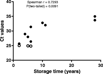
Methods: DNA in FFPE and FF tissue samples from heart, lungs, liver, kidneys, spleen and brain from patients with meningococcal shock and controls (lethal pneumococcal infection) stored at variable times, were isolated. The bacterial load of N. meningitidis DNA was analyzed using quantitative real-time PCR (qPCR) and primers for the capsule transport A (ctrA) gene (1 copy per N. meningitidis DNA). The human beta-hemoglobin (HBB) gene was quantified to evaluate effect of the storage times (2-28 years) and storage method in archived tissue.
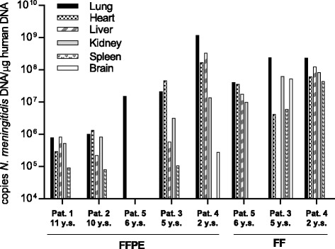
Results: N. meningitidis DNA was detected in FFPE and FF tissue samples from heart, lung, liver, kidney, and spleen in all patients with severe shock. In FFPE brain, N. meningitidis DNA was only detected in the patient with the highest concentration of LPS in the blood at admission to hospital. The highest levels of N. meningitidis DNA were found in heart tissue (median value 3.6 × 10⁷ copies N. meningitidis DNA/μg human DNA) and lung tissue (median value 3.1 × 10⁷ copies N. meningitidis DNA/μg human DNA) in all five patients. N. meningitidis DNA was not detectable in any of the tissue samples from two patients with clinical meningitis and the controls (pneumococcal infection). The quantity of HBB declined over time in FFPE tissue stored at room temperature, suggesting degradation of DNA.
Conclusions: High levels of N. meningitidis DNA were detected in the different tissue samples from meningococcal shock patients, particularly in the heart and lungs suggesting seeding and major proliferation of meningococci in these organs during the development of shock, probably contributing to the multiple organ failure. The age of archived tissue samples appear to have an impact on the amount of quantifiable N. meningitidis DNA.
期刊介绍:
BMC Clinical Pathology is an open access journal publishing original peer-reviewed research articles in all aspects of histopathology, haematology, clinical biochemistry, and medical microbiology (including virology, parasitology, and infection control). BMC Clinical Pathology (ISSN 1472-6890) is indexed/tracked/covered by PubMed, CAS, EMBASE, Scopus and Google Scholar.

 求助内容:
求助内容: 应助结果提醒方式:


