Meryem Rais, Amine Kessab, Zahra Sayad, Sanae El Mourabit, Redallah Zrarqi, Salma Benazzou, Malik Boulaadas, Nadia Cherradi
{"title":"孤立性纤维性肿瘤发生于腮腺:1例报告。","authors":"Meryem Rais, Amine Kessab, Zahra Sayad, Sanae El Mourabit, Redallah Zrarqi, Salma Benazzou, Malik Boulaadas, Nadia Cherradi","doi":"10.1186/s12907-017-0062-z","DOIUrl":null,"url":null,"abstract":"<p><strong>Background: </strong>Solitary fibrous tumor is an uncommon spindle cell neoplasm of unknown origin. It has been reported in many anatomic sites, with a rare occurrence in the head and neck region. Solitary fibrous tumors of the parotid gland are exceptional; their clinical and radiologic features are non specific, often mimicking more common salivary gland tumors. Pathologic examination and immunohistochemistry are required to make the correct diagnosis. The prognosis is favorable, with most tumors being benign, and complete surgical resection is the treatment of choice.</p><p><strong>Case presentation: </strong>We report the case of a 42-year-old man who presented with a painless mass involving the parotid gland. A parotidectomy was performed, and follow up was unremarkable. Gross examination showed a well circumscribed, firm tumor measuring 3,4 cm. Histologically, the tumor was composed of a spindle cell proliferation of variable cellularity, with staghorn vessels. A panel of immunohistochemical stains was performed, and confirmed the diagnosis of parotid gland solitary fibrous tumor.</p><p><strong>Conclusion: </strong>In this report we aim to increase awareness of this rare entity among clinicians and pathologists, and to emphasize the role of immunohistochemistry in confirming the diagnosis.</p>","PeriodicalId":35804,"journal":{"name":"BMC Clinical Pathology","volume":"17 ","pages":"22"},"PeriodicalIF":0.0000,"publicationDate":"2017-11-21","publicationTypes":"Journal Article","fieldsOfStudy":null,"isOpenAccess":false,"openAccessPdf":"https://sci-hub-pdf.com/10.1186/s12907-017-0062-z","citationCount":"10","resultStr":"{\"title\":\"Solitary fibrous tumor occurring in the parotid gland: a case report.\",\"authors\":\"Meryem Rais, Amine Kessab, Zahra Sayad, Sanae El Mourabit, Redallah Zrarqi, Salma Benazzou, Malik Boulaadas, Nadia Cherradi\",\"doi\":\"10.1186/s12907-017-0062-z\",\"DOIUrl\":null,\"url\":null,\"abstract\":\"<p><strong>Background: </strong>Solitary fibrous tumor is an uncommon spindle cell neoplasm of unknown origin. It has been reported in many anatomic sites, with a rare occurrence in the head and neck region. Solitary fibrous tumors of the parotid gland are exceptional; their clinical and radiologic features are non specific, often mimicking more common salivary gland tumors. Pathologic examination and immunohistochemistry are required to make the correct diagnosis. The prognosis is favorable, with most tumors being benign, and complete surgical resection is the treatment of choice.</p><p><strong>Case presentation: </strong>We report the case of a 42-year-old man who presented with a painless mass involving the parotid gland. A parotidectomy was performed, and follow up was unremarkable. Gross examination showed a well circumscribed, firm tumor measuring 3,4 cm. Histologically, the tumor was composed of a spindle cell proliferation of variable cellularity, with staghorn vessels. A panel of immunohistochemical stains was performed, and confirmed the diagnosis of parotid gland solitary fibrous tumor.</p><p><strong>Conclusion: </strong>In this report we aim to increase awareness of this rare entity among clinicians and pathologists, and to emphasize the role of immunohistochemistry in confirming the diagnosis.</p>\",\"PeriodicalId\":35804,\"journal\":{\"name\":\"BMC Clinical Pathology\",\"volume\":\"17 \",\"pages\":\"22\"},\"PeriodicalIF\":0.0000,\"publicationDate\":\"2017-11-21\",\"publicationTypes\":\"Journal Article\",\"fieldsOfStudy\":null,\"isOpenAccess\":false,\"openAccessPdf\":\"https://sci-hub-pdf.com/10.1186/s12907-017-0062-z\",\"citationCount\":\"10\",\"resultStr\":null,\"platform\":\"Semanticscholar\",\"paperid\":null,\"PeriodicalName\":\"BMC Clinical Pathology\",\"FirstCategoryId\":\"1085\",\"ListUrlMain\":\"https://doi.org/10.1186/s12907-017-0062-z\",\"RegionNum\":0,\"RegionCategory\":null,\"ArticlePicture\":[],\"TitleCN\":null,\"AbstractTextCN\":null,\"PMCID\":null,\"EPubDate\":\"2017/1/1 0:00:00\",\"PubModel\":\"eCollection\",\"JCR\":\"Q2\",\"JCRName\":\"Medicine\",\"Score\":null,\"Total\":0}","platform":"Semanticscholar","paperid":null,"PeriodicalName":"BMC Clinical Pathology","FirstCategoryId":"1085","ListUrlMain":"https://doi.org/10.1186/s12907-017-0062-z","RegionNum":0,"RegionCategory":null,"ArticlePicture":[],"TitleCN":null,"AbstractTextCN":null,"PMCID":null,"EPubDate":"2017/1/1 0:00:00","PubModel":"eCollection","JCR":"Q2","JCRName":"Medicine","Score":null,"Total":0}
Solitary fibrous tumor occurring in the parotid gland: a case report.
Background: Solitary fibrous tumor is an uncommon spindle cell neoplasm of unknown origin. It has been reported in many anatomic sites, with a rare occurrence in the head and neck region. Solitary fibrous tumors of the parotid gland are exceptional; their clinical and radiologic features are non specific, often mimicking more common salivary gland tumors. Pathologic examination and immunohistochemistry are required to make the correct diagnosis. The prognosis is favorable, with most tumors being benign, and complete surgical resection is the treatment of choice.
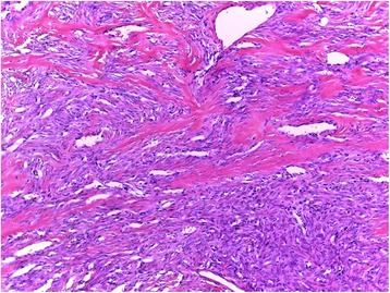
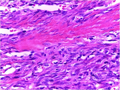
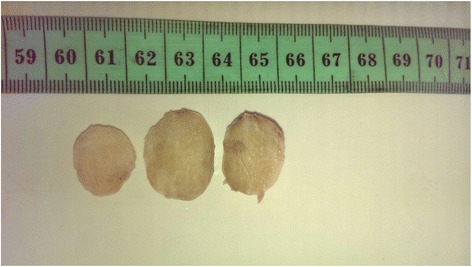
Case presentation: We report the case of a 42-year-old man who presented with a painless mass involving the parotid gland. A parotidectomy was performed, and follow up was unremarkable. Gross examination showed a well circumscribed, firm tumor measuring 3,4 cm. Histologically, the tumor was composed of a spindle cell proliferation of variable cellularity, with staghorn vessels. A panel of immunohistochemical stains was performed, and confirmed the diagnosis of parotid gland solitary fibrous tumor.
Conclusion: In this report we aim to increase awareness of this rare entity among clinicians and pathologists, and to emphasize the role of immunohistochemistry in confirming the diagnosis.
期刊介绍:
BMC Clinical Pathology is an open access journal publishing original peer-reviewed research articles in all aspects of histopathology, haematology, clinical biochemistry, and medical microbiology (including virology, parasitology, and infection control). BMC Clinical Pathology (ISSN 1472-6890) is indexed/tracked/covered by PubMed, CAS, EMBASE, Scopus and Google Scholar.

 求助内容:
求助内容: 应助结果提醒方式:


