Sasapin G Prakalapakorn, Laura A Vickers, Rolando Estrada, Sharon F Freedman, Carlo Tomasi, Sina Farsiu, David K Wallace
{"title":"利用图像融合方法提高ROPtool后极血管分析的效率和可追溯性。","authors":"Sasapin G Prakalapakorn, Laura A Vickers, Rolando Estrada, Sharon F Freedman, Carlo Tomasi, Sina Farsiu, David K Wallace","doi":"10.2174/1874364101711010143","DOIUrl":null,"url":null,"abstract":"<p><strong>Background: </strong>The diagnosis of plus disease in retinopathy of prematurity (ROP) largely determines the need for treatment; however, this diagnosis is subjective. To make the diagnosis of plus disease more objective, semi-automated computer programs (<i>e.g.</i> ROPtool) have been created to quantify vascular dilation and tortuosity. ROPtool can accurately analyze blood vessels only in images with very good quality, but many still images captured by indirect ophthalmoscopy have insufficient image quality for ROPtool analysis.</p><p><strong>Purpose: </strong>To evaluate the ability of an image fusion methodology (robust mosaicing) to increase the efficiency and traceability of posterior pole vessel analysis by ROPtool.</p><p><strong>Materials and methodology: </strong>We retrospectively reviewed video indirect ophthalmoscopy images acquired during routine ROP examinations and selected the best unenhanced still image from the video for each infant. Robust mosaicing was used to create an enhanced mosaic image from the same video for each eye. We evaluated the time required for ROPtool analysis as well as ROPtool's ability to analyze vessels in enhanced vs. unenhanced images.</p><p><strong>Results: </strong>We included 39 eyes of 39 infants. ROPtool analysis was faster (125 vs. 152 seconds; <i>p</i>=0.02) in enhanced vs. unenhanced images, respectively. ROPtool was able to trace retinal vessels in more quadrants (143/156, 92% vs 115/156, 74%; p=0.16) in enhanced mosaic vs. unenhanced still images, respectively and in more overall (38/39, 97% vs. 34/39, 87%; p=0.07) enhanced mosaic vs. unenhanced still images, respectively.</p><p><strong>Conclusion: </strong>Retinal image enhancement using robust mosaicing advances efforts to automate grading of posterior pole disease in ROP.</p>","PeriodicalId":512318,"journal":{"name":"The Open Ophthalmology Journal","volume":"11 ","pages":"143-151"},"PeriodicalIF":0.0000,"publicationDate":"2017-06-29","publicationTypes":"Journal Article","fieldsOfStudy":null,"isOpenAccess":false,"openAccessPdf":"https://www.ncbi.nlm.nih.gov/pmc/articles/PMC5510566/pdf/","citationCount":"1","resultStr":"{\"title\":\"Using an Image Fusion Methodology to Improve Efficiency and Traceability of Posterior Pole Vessel Analysis by ROPtool.\",\"authors\":\"Sasapin G Prakalapakorn, Laura A Vickers, Rolando Estrada, Sharon F Freedman, Carlo Tomasi, Sina Farsiu, David K Wallace\",\"doi\":\"10.2174/1874364101711010143\",\"DOIUrl\":null,\"url\":null,\"abstract\":\"<p><strong>Background: </strong>The diagnosis of plus disease in retinopathy of prematurity (ROP) largely determines the need for treatment; however, this diagnosis is subjective. To make the diagnosis of plus disease more objective, semi-automated computer programs (<i>e.g.</i> ROPtool) have been created to quantify vascular dilation and tortuosity. ROPtool can accurately analyze blood vessels only in images with very good quality, but many still images captured by indirect ophthalmoscopy have insufficient image quality for ROPtool analysis.</p><p><strong>Purpose: </strong>To evaluate the ability of an image fusion methodology (robust mosaicing) to increase the efficiency and traceability of posterior pole vessel analysis by ROPtool.</p><p><strong>Materials and methodology: </strong>We retrospectively reviewed video indirect ophthalmoscopy images acquired during routine ROP examinations and selected the best unenhanced still image from the video for each infant. Robust mosaicing was used to create an enhanced mosaic image from the same video for each eye. We evaluated the time required for ROPtool analysis as well as ROPtool's ability to analyze vessels in enhanced vs. unenhanced images.</p><p><strong>Results: </strong>We included 39 eyes of 39 infants. ROPtool analysis was faster (125 vs. 152 seconds; <i>p</i>=0.02) in enhanced vs. unenhanced images, respectively. ROPtool was able to trace retinal vessels in more quadrants (143/156, 92% vs 115/156, 74%; p=0.16) in enhanced mosaic vs. unenhanced still images, respectively and in more overall (38/39, 97% vs. 34/39, 87%; p=0.07) enhanced mosaic vs. unenhanced still images, respectively.</p><p><strong>Conclusion: </strong>Retinal image enhancement using robust mosaicing advances efforts to automate grading of posterior pole disease in ROP.</p>\",\"PeriodicalId\":512318,\"journal\":{\"name\":\"The Open Ophthalmology Journal\",\"volume\":\"11 \",\"pages\":\"143-151\"},\"PeriodicalIF\":0.0000,\"publicationDate\":\"2017-06-29\",\"publicationTypes\":\"Journal Article\",\"fieldsOfStudy\":null,\"isOpenAccess\":false,\"openAccessPdf\":\"https://www.ncbi.nlm.nih.gov/pmc/articles/PMC5510566/pdf/\",\"citationCount\":\"1\",\"resultStr\":null,\"platform\":\"Semanticscholar\",\"paperid\":null,\"PeriodicalName\":\"The Open Ophthalmology Journal\",\"FirstCategoryId\":\"1085\",\"ListUrlMain\":\"https://doi.org/10.2174/1874364101711010143\",\"RegionNum\":0,\"RegionCategory\":null,\"ArticlePicture\":[],\"TitleCN\":null,\"AbstractTextCN\":null,\"PMCID\":null,\"EPubDate\":\"2017/1/1 0:00:00\",\"PubModel\":\"eCollection\",\"JCR\":\"\",\"JCRName\":\"\",\"Score\":null,\"Total\":0}","platform":"Semanticscholar","paperid":null,"PeriodicalName":"The Open Ophthalmology Journal","FirstCategoryId":"1085","ListUrlMain":"https://doi.org/10.2174/1874364101711010143","RegionNum":0,"RegionCategory":null,"ArticlePicture":[],"TitleCN":null,"AbstractTextCN":null,"PMCID":null,"EPubDate":"2017/1/1 0:00:00","PubModel":"eCollection","JCR":"","JCRName":"","Score":null,"Total":0}
引用次数: 1
摘要
背景:早产儿视网膜病变(ROP)加病的诊断在很大程度上决定了治疗的需要;然而,这种诊断是主观的。为了使正性疾病的诊断更加客观,已经创建了半自动计算机程序(例如ROPtool)来量化血管扩张和扭曲。ROPtool只能对质量非常好的图像进行准确的血管分析,而许多间接检眼镜拍摄的静止图像质量不足,无法进行ROPtool分析。目的:评估图像融合方法(鲁棒拼接)提高ROPtool后极血管分析效率和可追溯性的能力。材料和方法:我们回顾性地回顾了在常规ROP检查中获得的视频间接检眼镜图像,并从视频中为每个婴儿选择了最佳的未增强静态图像。鲁棒拼接用于从同一视频中为每只眼睛创建增强的马赛克图像。我们评估了ROPtool分析所需的时间,以及ROPtool在增强和未增强图像中分析血管的能力。结果:我们纳入了39例婴儿的39只眼睛。ROPtool分析更快(125秒vs 152秒);P =0.02)。ROPtool能够追踪更多象限的视网膜血管(143/156,92% vs 115/156, 74%;P =0.16),在增强的马赛克图像和未增强的静止图像中分别,在更全面的情况下(38/ 39,97% vs. 34/ 39,87%;P =0.07)增强的马赛克与未增强的静止图像。结论:采用鲁棒拼接技术增强视网膜图像,有助于ROP后极病变的自动分级。
Using an Image Fusion Methodology to Improve Efficiency and Traceability of Posterior Pole Vessel Analysis by ROPtool.
Background: The diagnosis of plus disease in retinopathy of prematurity (ROP) largely determines the need for treatment; however, this diagnosis is subjective. To make the diagnosis of plus disease more objective, semi-automated computer programs (e.g. ROPtool) have been created to quantify vascular dilation and tortuosity. ROPtool can accurately analyze blood vessels only in images with very good quality, but many still images captured by indirect ophthalmoscopy have insufficient image quality for ROPtool analysis.
Purpose: To evaluate the ability of an image fusion methodology (robust mosaicing) to increase the efficiency and traceability of posterior pole vessel analysis by ROPtool.
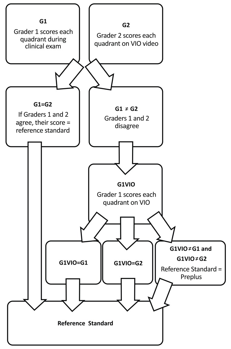
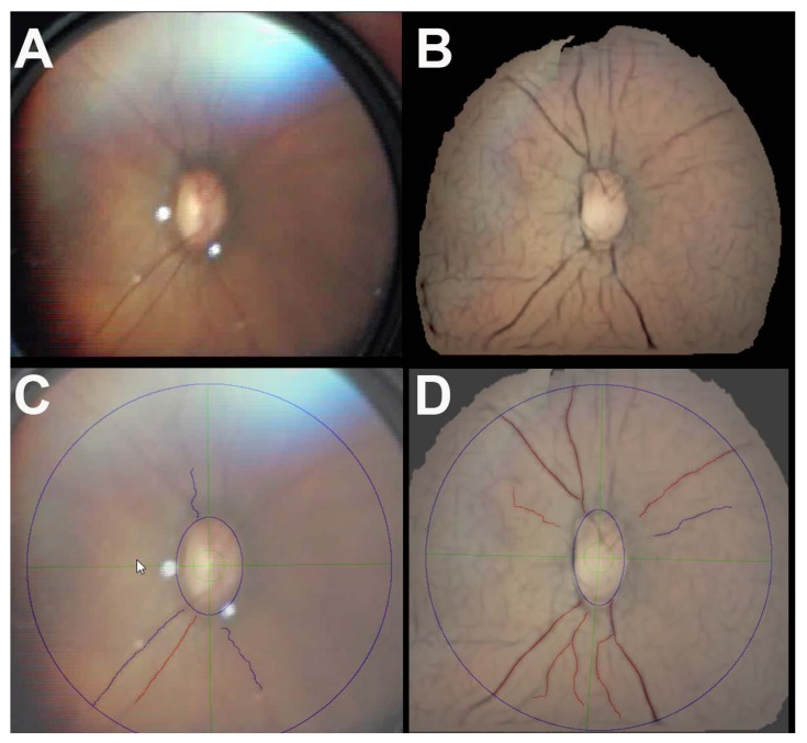
Materials and methodology: We retrospectively reviewed video indirect ophthalmoscopy images acquired during routine ROP examinations and selected the best unenhanced still image from the video for each infant. Robust mosaicing was used to create an enhanced mosaic image from the same video for each eye. We evaluated the time required for ROPtool analysis as well as ROPtool's ability to analyze vessels in enhanced vs. unenhanced images.
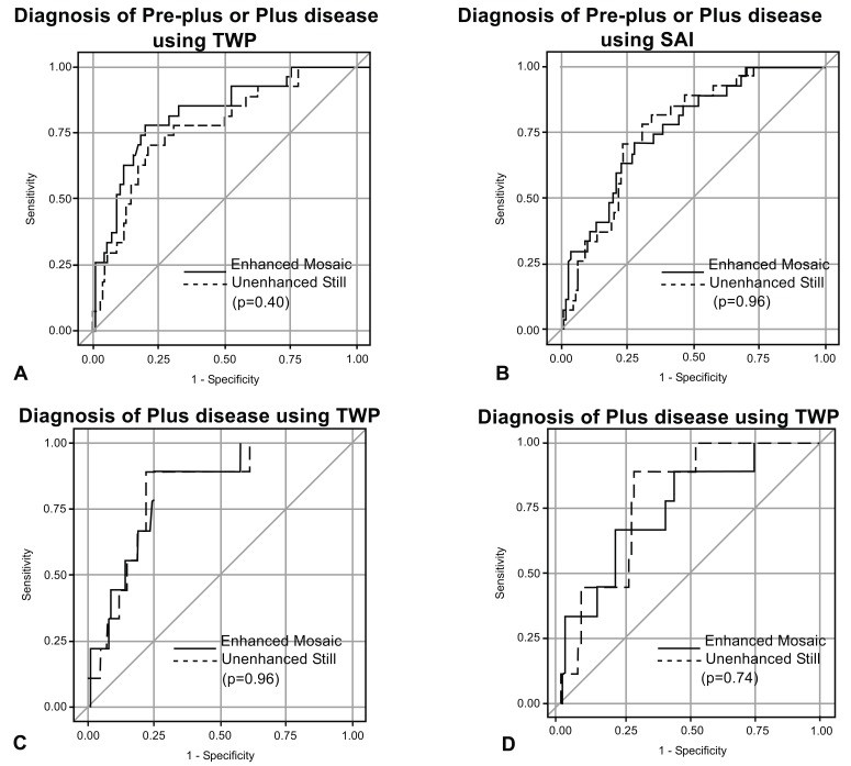
Results: We included 39 eyes of 39 infants. ROPtool analysis was faster (125 vs. 152 seconds; p=0.02) in enhanced vs. unenhanced images, respectively. ROPtool was able to trace retinal vessels in more quadrants (143/156, 92% vs 115/156, 74%; p=0.16) in enhanced mosaic vs. unenhanced still images, respectively and in more overall (38/39, 97% vs. 34/39, 87%; p=0.07) enhanced mosaic vs. unenhanced still images, respectively.
Conclusion: Retinal image enhancement using robust mosaicing advances efforts to automate grading of posterior pole disease in ROP.

 求助内容:
求助内容: 应助结果提醒方式:


