{"title":"光学相干断层扫描(OCT)诊断色素性视网膜炎个案研究。","authors":"Melita Adilovic, Emira Ignjatic, Arnes Cabric","doi":"10.5455/aim.2022.30.329-333","DOIUrl":null,"url":null,"abstract":"<p><strong>Background: </strong>Retinitis pigmentosa (RP) is a set of inherited rod-cone degenerative diseases that clinically presents with similar signs and symptoms. Mutations in one of more than 70 genes are involved. Patients will commonly present with bone-spicule pigment formation, waxy optic nerve pallor, and attenuated blood vessels in the posterior pole.Symptoms often begin with progressive night blindness, mid-peripheral visual field defects, and eventual tunnel vision. Central vision loss will ultimately occur following loss of rod function. Complete blindness is uncommon.</p><p><strong>Objective: </strong>The aim of this article is to present two cases of retinitis pigmentosa (mother and daughter) trough optalmologic exams in our clinic. The next aim it to show how to menage a low vision service and to treat cystoid macular oedema as a complication of retinitis pigmentosa.</p><p><strong>Methods: </strong>All medical reports are shown in this article. Every diagnostic tool as well as report is a part from our archived history of the patients and has been throughly analysed. We also reviewed available literature using the key words retinitis pigmentosa, cystoid macular oedema, gene therapy.</p><p><strong>Case presentation: </strong>A 38 year old female patient for a low vision consultation. The patient was legally blind secondary to retinitis pigmentosa, which was diagnosed in her late 20s. She reported gradually progressive hazy central vision and decreasing peripheral vision in both eyes as well as severe night blindness. Other than the diagnosis of retinitis pigmentosa in both eyes,the patient had no other remarkable ocular conditions. Findings at that visit included unaided distance visual acuities VOD: 0,04 VOS: 0,06. Pupils were round with brisk responses. Extraocular muscle motility was full in both eyes. Confrontation methode visual fields were noted as temporal loss in the right eye and superior and temporal loss in the left eye. The perimetry test could not be performed due to the lack of correspondece of the patient even after a couple repetitions of the perimetry. She had normal ocular adnexa and quiet lids, conjunctiva, and sclera in both eyes. Corneas in both eyes were noted as clear epithelium, clear stroma, and clear endothelium. Anterior chambers had normal depth, iris with no pathological findings in both eyes; lens incipient sclerotic. Intraocular pressures were noted as 22 mmHg in both eyes with Icare, 21mmHg and 19 mmHg with aplanation tonometry; pahimetry corretional factor was +1 on both eyes. The vitreous was clear in both eyes. Both optic nerves were measured as 0.4 cup-to-disc ratios with no disc edema, disc hemorrhages, notching, or thinning noted.Waxy disc pallor and attenuated blood vessels were observed in both eyes. The macula in both eyes had retinal pigment epithelium (RPE) changes with no edema or hemorrhages. Bone spicule changes were noted 360 in the periphery of both eyes with no holes or tears(Figure 1a+1b+1c+1d).</p><p><strong>Conclusion: </strong>We presented two cases of retinitis pigmentosa - the mother with diagnosed RP more than 15 years ago in need for low vision rehabilitation service and the daughter that got diagnosed after our initial examination and with complications in visual impairment through cystoid macular oedema.</p>","PeriodicalId":7074,"journal":{"name":"Acta Informatica Medica","volume":" ","pages":"329-333"},"PeriodicalIF":0.0000,"publicationDate":"2022-12-01","publicationTypes":"Journal Article","fieldsOfStudy":null,"isOpenAccess":false,"openAccessPdf":"https://ftp.ncbi.nlm.nih.gov/pub/pmc/oa_pdf/56/66/AIM-30-329.PMC9665416.pdf","citationCount":"1","resultStr":"{\"title\":\"Optical Coherence Tomography (OCT) Diagnostic of Retinitis Pigmentosa - Case Study.\",\"authors\":\"Melita Adilovic, Emira Ignjatic, Arnes Cabric\",\"doi\":\"10.5455/aim.2022.30.329-333\",\"DOIUrl\":null,\"url\":null,\"abstract\":\"<p><strong>Background: </strong>Retinitis pigmentosa (RP) is a set of inherited rod-cone degenerative diseases that clinically presents with similar signs and symptoms. Mutations in one of more than 70 genes are involved. Patients will commonly present with bone-spicule pigment formation, waxy optic nerve pallor, and attenuated blood vessels in the posterior pole.Symptoms often begin with progressive night blindness, mid-peripheral visual field defects, and eventual tunnel vision. Central vision loss will ultimately occur following loss of rod function. Complete blindness is uncommon.</p><p><strong>Objective: </strong>The aim of this article is to present two cases of retinitis pigmentosa (mother and daughter) trough optalmologic exams in our clinic. The next aim it to show how to menage a low vision service and to treat cystoid macular oedema as a complication of retinitis pigmentosa.</p><p><strong>Methods: </strong>All medical reports are shown in this article. Every diagnostic tool as well as report is a part from our archived history of the patients and has been throughly analysed. We also reviewed available literature using the key words retinitis pigmentosa, cystoid macular oedema, gene therapy.</p><p><strong>Case presentation: </strong>A 38 year old female patient for a low vision consultation. The patient was legally blind secondary to retinitis pigmentosa, which was diagnosed in her late 20s. She reported gradually progressive hazy central vision and decreasing peripheral vision in both eyes as well as severe night blindness. Other than the diagnosis of retinitis pigmentosa in both eyes,the patient had no other remarkable ocular conditions. Findings at that visit included unaided distance visual acuities VOD: 0,04 VOS: 0,06. Pupils were round with brisk responses. Extraocular muscle motility was full in both eyes. Confrontation methode visual fields were noted as temporal loss in the right eye and superior and temporal loss in the left eye. The perimetry test could not be performed due to the lack of correspondece of the patient even after a couple repetitions of the perimetry. She had normal ocular adnexa and quiet lids, conjunctiva, and sclera in both eyes. Corneas in both eyes were noted as clear epithelium, clear stroma, and clear endothelium. Anterior chambers had normal depth, iris with no pathological findings in both eyes; lens incipient sclerotic. Intraocular pressures were noted as 22 mmHg in both eyes with Icare, 21mmHg and 19 mmHg with aplanation tonometry; pahimetry corretional factor was +1 on both eyes. The vitreous was clear in both eyes. Both optic nerves were measured as 0.4 cup-to-disc ratios with no disc edema, disc hemorrhages, notching, or thinning noted.Waxy disc pallor and attenuated blood vessels were observed in both eyes. The macula in both eyes had retinal pigment epithelium (RPE) changes with no edema or hemorrhages. Bone spicule changes were noted 360 in the periphery of both eyes with no holes or tears(Figure 1a+1b+1c+1d).</p><p><strong>Conclusion: </strong>We presented two cases of retinitis pigmentosa - the mother with diagnosed RP more than 15 years ago in need for low vision rehabilitation service and the daughter that got diagnosed after our initial examination and with complications in visual impairment through cystoid macular oedema.</p>\",\"PeriodicalId\":7074,\"journal\":{\"name\":\"Acta Informatica Medica\",\"volume\":\" \",\"pages\":\"329-333\"},\"PeriodicalIF\":0.0000,\"publicationDate\":\"2022-12-01\",\"publicationTypes\":\"Journal Article\",\"fieldsOfStudy\":null,\"isOpenAccess\":false,\"openAccessPdf\":\"https://ftp.ncbi.nlm.nih.gov/pub/pmc/oa_pdf/56/66/AIM-30-329.PMC9665416.pdf\",\"citationCount\":\"1\",\"resultStr\":null,\"platform\":\"Semanticscholar\",\"paperid\":null,\"PeriodicalName\":\"Acta Informatica Medica\",\"FirstCategoryId\":\"1085\",\"ListUrlMain\":\"https://doi.org/10.5455/aim.2022.30.329-333\",\"RegionNum\":0,\"RegionCategory\":null,\"ArticlePicture\":[],\"TitleCN\":null,\"AbstractTextCN\":null,\"PMCID\":null,\"EPubDate\":\"\",\"PubModel\":\"\",\"JCR\":\"Q2\",\"JCRName\":\"Medicine\",\"Score\":null,\"Total\":0}","platform":"Semanticscholar","paperid":null,"PeriodicalName":"Acta Informatica Medica","FirstCategoryId":"1085","ListUrlMain":"https://doi.org/10.5455/aim.2022.30.329-333","RegionNum":0,"RegionCategory":null,"ArticlePicture":[],"TitleCN":null,"AbstractTextCN":null,"PMCID":null,"EPubDate":"","PubModel":"","JCR":"Q2","JCRName":"Medicine","Score":null,"Total":0}
Optical Coherence Tomography (OCT) Diagnostic of Retinitis Pigmentosa - Case Study.
Background: Retinitis pigmentosa (RP) is a set of inherited rod-cone degenerative diseases that clinically presents with similar signs and symptoms. Mutations in one of more than 70 genes are involved. Patients will commonly present with bone-spicule pigment formation, waxy optic nerve pallor, and attenuated blood vessels in the posterior pole.Symptoms often begin with progressive night blindness, mid-peripheral visual field defects, and eventual tunnel vision. Central vision loss will ultimately occur following loss of rod function. Complete blindness is uncommon.
Objective: The aim of this article is to present two cases of retinitis pigmentosa (mother and daughter) trough optalmologic exams in our clinic. The next aim it to show how to menage a low vision service and to treat cystoid macular oedema as a complication of retinitis pigmentosa.
Methods: All medical reports are shown in this article. Every diagnostic tool as well as report is a part from our archived history of the patients and has been throughly analysed. We also reviewed available literature using the key words retinitis pigmentosa, cystoid macular oedema, gene therapy.
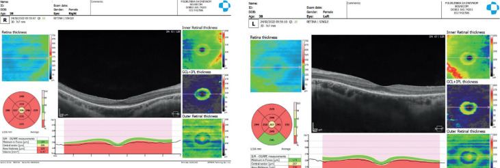
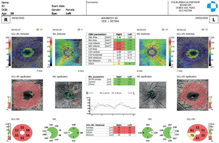
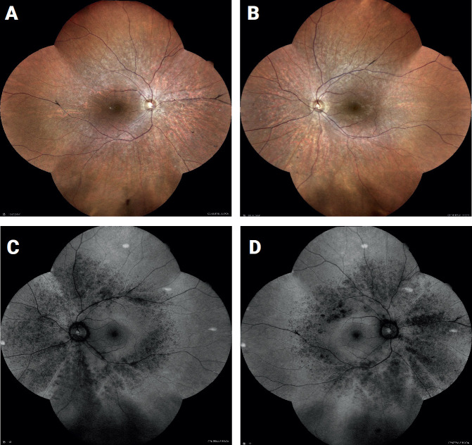
Case presentation: A 38 year old female patient for a low vision consultation. The patient was legally blind secondary to retinitis pigmentosa, which was diagnosed in her late 20s. She reported gradually progressive hazy central vision and decreasing peripheral vision in both eyes as well as severe night blindness. Other than the diagnosis of retinitis pigmentosa in both eyes,the patient had no other remarkable ocular conditions. Findings at that visit included unaided distance visual acuities VOD: 0,04 VOS: 0,06. Pupils were round with brisk responses. Extraocular muscle motility was full in both eyes. Confrontation methode visual fields were noted as temporal loss in the right eye and superior and temporal loss in the left eye. The perimetry test could not be performed due to the lack of correspondece of the patient even after a couple repetitions of the perimetry. She had normal ocular adnexa and quiet lids, conjunctiva, and sclera in both eyes. Corneas in both eyes were noted as clear epithelium, clear stroma, and clear endothelium. Anterior chambers had normal depth, iris with no pathological findings in both eyes; lens incipient sclerotic. Intraocular pressures were noted as 22 mmHg in both eyes with Icare, 21mmHg and 19 mmHg with aplanation tonometry; pahimetry corretional factor was +1 on both eyes. The vitreous was clear in both eyes. Both optic nerves were measured as 0.4 cup-to-disc ratios with no disc edema, disc hemorrhages, notching, or thinning noted.Waxy disc pallor and attenuated blood vessels were observed in both eyes. The macula in both eyes had retinal pigment epithelium (RPE) changes with no edema or hemorrhages. Bone spicule changes were noted 360 in the periphery of both eyes with no holes or tears(Figure 1a+1b+1c+1d).
Conclusion: We presented two cases of retinitis pigmentosa - the mother with diagnosed RP more than 15 years ago in need for low vision rehabilitation service and the daughter that got diagnosed after our initial examination and with complications in visual impairment through cystoid macular oedema.

 求助内容:
求助内容: 应助结果提醒方式:


