{"title":"瑞典年轻人视网膜中央静脉闭塞:病例报告和文献回顾。","authors":"Elisabeth Wittström","doi":"10.2174/1874364101711010089","DOIUrl":null,"url":null,"abstract":"<p><strong>Purpose: </strong>To investigate associated systemic diseases, other conditions, visual outcome, ocular complications and treatment in Swedish patients younger than 50 years with central retinal vein occlusion (CRVO) and reviewing the literature.</p><p><strong>Methods: </strong>Twenty-two patients with CRVO, younger than 50 years, were examined with full-field electroretinography (ERG) within 3 months after a thrombotic event, or were periodically examined and were observed for at least 6 months. In 18 of these patients, the initial retinal ischemia was studied using the cone b-wave implicit time in the 30 Hz flicker ERG. Fifteen patients also underwent fluorescein angiography. Optical coherence tomography (OCT) was performed in 14 patients. The patients studied were divided into two groups, non-ischemic and ischemic, which were compared. All patients underwent ocular and systemic examination, as well as complete screening for thrombophilic risk factors.</p><p><strong>Results: </strong>Of the 22 patients, 15 had non-ischemic type of CRVO and 7 the ischemic type. Patients with non-ischemic CRVO showed significantly improved visual acuity (VA) at the final examination (p=0.006). Patients with ischemic CRVO showed no significant reduction in VA at the final examination (p=0.225). Systemic hypertension (27% in non-ischemic CRVO and 29% in ischemic CRVO) was the most prevalent systemic risk factor for CRVO. The mean central foveal thickness (CFT) decreased significantly from 402.3±136.2 (µm) at the initial examination to 243.8±48.1 (µm) at the final examination in the non-ischemic group (p=0.005). The mean initial CFT was 444.5±186.1 (µm) in the ischemic CRVO group, which decreased to 211.5±20.2 (µm) at the final visit (p=0.068). Pigment dispersion syndrome (PDS)/pigmentary glaucoma (PG), ocular hypertension and dehydration were equally frequent; four patients each (18%) out of 22. The clinical course of 4 younger patients with PDS/PG are described.</p><p><strong>Conclusion: </strong>The patients with non-ischemic CRVO showed significantly improved VA and significantly decreased CFT at the final examination. Systemic hypertension was the most prevalent risk factor for CRVO. Younger adults with CRVO also had a high prevalence of PDS/PG, ocular hypertension and dehydration. This study highlights the importance of careful IOP monitoring, and the need to investigate possible PDS/PG and to obtain an accurate history of the patient including alcohol intake and intense exercise.</p>","PeriodicalId":512318,"journal":{"name":"The Open Ophthalmology Journal","volume":"11 ","pages":"89-102"},"PeriodicalIF":0.0000,"publicationDate":"2017-05-22","publicationTypes":"Journal Article","fieldsOfStudy":null,"isOpenAccess":false,"openAccessPdf":"https://www.ncbi.nlm.nih.gov/pmc/articles/PMC5447937/pdf/","citationCount":"9","resultStr":"{\"title\":\"Central Retinal Vein Occlusion in Younger Swedish Adults: Case Reports and Review of the Literature.\",\"authors\":\"Elisabeth Wittström\",\"doi\":\"10.2174/1874364101711010089\",\"DOIUrl\":null,\"url\":null,\"abstract\":\"<p><strong>Purpose: </strong>To investigate associated systemic diseases, other conditions, visual outcome, ocular complications and treatment in Swedish patients younger than 50 years with central retinal vein occlusion (CRVO) and reviewing the literature.</p><p><strong>Methods: </strong>Twenty-two patients with CRVO, younger than 50 years, were examined with full-field electroretinography (ERG) within 3 months after a thrombotic event, or were periodically examined and were observed for at least 6 months. In 18 of these patients, the initial retinal ischemia was studied using the cone b-wave implicit time in the 30 Hz flicker ERG. Fifteen patients also underwent fluorescein angiography. Optical coherence tomography (OCT) was performed in 14 patients. The patients studied were divided into two groups, non-ischemic and ischemic, which were compared. All patients underwent ocular and systemic examination, as well as complete screening for thrombophilic risk factors.</p><p><strong>Results: </strong>Of the 22 patients, 15 had non-ischemic type of CRVO and 7 the ischemic type. Patients with non-ischemic CRVO showed significantly improved visual acuity (VA) at the final examination (p=0.006). Patients with ischemic CRVO showed no significant reduction in VA at the final examination (p=0.225). Systemic hypertension (27% in non-ischemic CRVO and 29% in ischemic CRVO) was the most prevalent systemic risk factor for CRVO. The mean central foveal thickness (CFT) decreased significantly from 402.3±136.2 (µm) at the initial examination to 243.8±48.1 (µm) at the final examination in the non-ischemic group (p=0.005). The mean initial CFT was 444.5±186.1 (µm) in the ischemic CRVO group, which decreased to 211.5±20.2 (µm) at the final visit (p=0.068). Pigment dispersion syndrome (PDS)/pigmentary glaucoma (PG), ocular hypertension and dehydration were equally frequent; four patients each (18%) out of 22. The clinical course of 4 younger patients with PDS/PG are described.</p><p><strong>Conclusion: </strong>The patients with non-ischemic CRVO showed significantly improved VA and significantly decreased CFT at the final examination. Systemic hypertension was the most prevalent risk factor for CRVO. Younger adults with CRVO also had a high prevalence of PDS/PG, ocular hypertension and dehydration. This study highlights the importance of careful IOP monitoring, and the need to investigate possible PDS/PG and to obtain an accurate history of the patient including alcohol intake and intense exercise.</p>\",\"PeriodicalId\":512318,\"journal\":{\"name\":\"The Open Ophthalmology Journal\",\"volume\":\"11 \",\"pages\":\"89-102\"},\"PeriodicalIF\":0.0000,\"publicationDate\":\"2017-05-22\",\"publicationTypes\":\"Journal Article\",\"fieldsOfStudy\":null,\"isOpenAccess\":false,\"openAccessPdf\":\"https://www.ncbi.nlm.nih.gov/pmc/articles/PMC5447937/pdf/\",\"citationCount\":\"9\",\"resultStr\":null,\"platform\":\"Semanticscholar\",\"paperid\":null,\"PeriodicalName\":\"The Open Ophthalmology Journal\",\"FirstCategoryId\":\"1085\",\"ListUrlMain\":\"https://doi.org/10.2174/1874364101711010089\",\"RegionNum\":0,\"RegionCategory\":null,\"ArticlePicture\":[],\"TitleCN\":null,\"AbstractTextCN\":null,\"PMCID\":null,\"EPubDate\":\"2017/1/1 0:00:00\",\"PubModel\":\"eCollection\",\"JCR\":\"\",\"JCRName\":\"\",\"Score\":null,\"Total\":0}","platform":"Semanticscholar","paperid":null,"PeriodicalName":"The Open Ophthalmology Journal","FirstCategoryId":"1085","ListUrlMain":"https://doi.org/10.2174/1874364101711010089","RegionNum":0,"RegionCategory":null,"ArticlePicture":[],"TitleCN":null,"AbstractTextCN":null,"PMCID":null,"EPubDate":"2017/1/1 0:00:00","PubModel":"eCollection","JCR":"","JCRName":"","Score":null,"Total":0}
Central Retinal Vein Occlusion in Younger Swedish Adults: Case Reports and Review of the Literature.
Purpose: To investigate associated systemic diseases, other conditions, visual outcome, ocular complications and treatment in Swedish patients younger than 50 years with central retinal vein occlusion (CRVO) and reviewing the literature.
Methods: Twenty-two patients with CRVO, younger than 50 years, were examined with full-field electroretinography (ERG) within 3 months after a thrombotic event, or were periodically examined and were observed for at least 6 months. In 18 of these patients, the initial retinal ischemia was studied using the cone b-wave implicit time in the 30 Hz flicker ERG. Fifteen patients also underwent fluorescein angiography. Optical coherence tomography (OCT) was performed in 14 patients. The patients studied were divided into two groups, non-ischemic and ischemic, which were compared. All patients underwent ocular and systemic examination, as well as complete screening for thrombophilic risk factors.
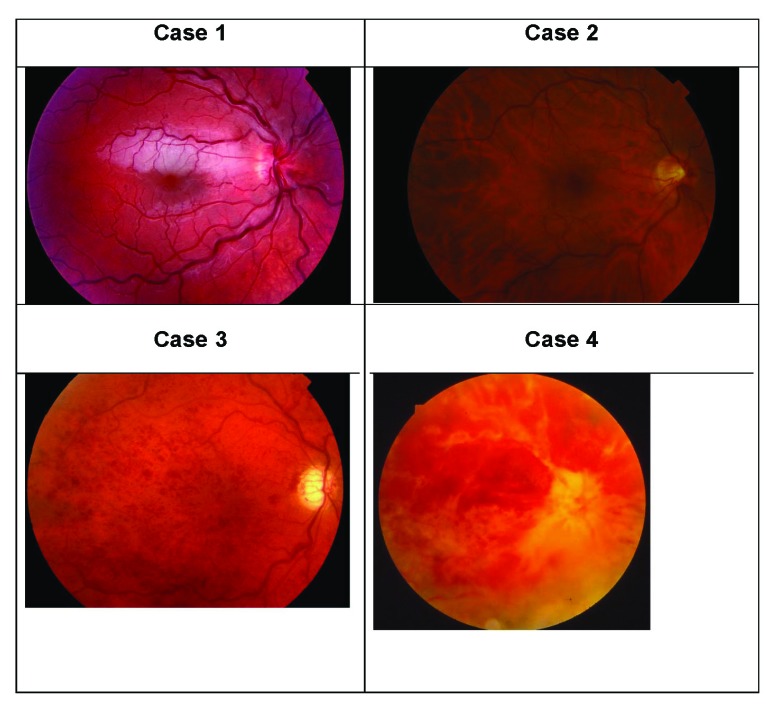
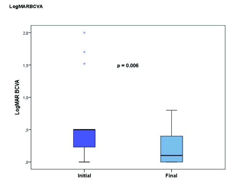
Results: Of the 22 patients, 15 had non-ischemic type of CRVO and 7 the ischemic type. Patients with non-ischemic CRVO showed significantly improved visual acuity (VA) at the final examination (p=0.006). Patients with ischemic CRVO showed no significant reduction in VA at the final examination (p=0.225). Systemic hypertension (27% in non-ischemic CRVO and 29% in ischemic CRVO) was the most prevalent systemic risk factor for CRVO. The mean central foveal thickness (CFT) decreased significantly from 402.3±136.2 (µm) at the initial examination to 243.8±48.1 (µm) at the final examination in the non-ischemic group (p=0.005). The mean initial CFT was 444.5±186.1 (µm) in the ischemic CRVO group, which decreased to 211.5±20.2 (µm) at the final visit (p=0.068). Pigment dispersion syndrome (PDS)/pigmentary glaucoma (PG), ocular hypertension and dehydration were equally frequent; four patients each (18%) out of 22. The clinical course of 4 younger patients with PDS/PG are described.
Conclusion: The patients with non-ischemic CRVO showed significantly improved VA and significantly decreased CFT at the final examination. Systemic hypertension was the most prevalent risk factor for CRVO. Younger adults with CRVO also had a high prevalence of PDS/PG, ocular hypertension and dehydration. This study highlights the importance of careful IOP monitoring, and the need to investigate possible PDS/PG and to obtain an accurate history of the patient including alcohol intake and intense exercise.

 求助内容:
求助内容: 应助结果提醒方式:


