{"title":"基于板束几何的多色x射线荧光计算机断层扫描蒙特卡罗模拟。","authors":"Shanghai Jiang, Peng He, Luzhen Deng, Mianyi Chen, Biao Wei","doi":"10.1155/2017/7916260","DOIUrl":null,"url":null,"abstract":"<p><p>X-ray fluorescence computed tomography (XFCT) based on sheet beam can save a huge amount of time to obtain a whole set of projections using synchrotron. However, it is clearly unpractical for most biomedical research laboratories. In this paper, polychromatic X-ray fluorescence computed tomography with sheet-beam geometry is tested by Monte Carlo simulation. First, two phantoms (<i>A</i> and <i>B</i>) filled with PMMA are used to simulate imaging process through GEANT 4. Phantom <i>A</i> contains several GNP-loaded regions with the same size (10 mm) in height and diameter but different Au weight concentration ranging from 0.3% to 1.8%. Phantom <i>B</i> contains twelve GNP-loaded regions with the same Au weight concentration (1.6%) but different diameter ranging from 1 mm to 9 mm. Second, discretized presentation of imaging model is established to reconstruct more accurate XFCT images. Third, XFCT images of phantoms <i>A</i> and <i>B</i> are reconstructed by filter back-projection (FBP) and maximum likelihood expectation maximization (MLEM) with and without correction, respectively. Contrast-to-noise ratio (CNR) is calculated to evaluate all the reconstructed images. Our results show that it is feasible for sheet-beam XFCT system based on polychromatic X-ray source and the discretized imaging model can be used to reconstruct more accurate images.</p>","PeriodicalId":47063,"journal":{"name":"International Journal of Biomedical Imaging","volume":"2017 ","pages":"7916260"},"PeriodicalIF":1.3000,"publicationDate":"2017-01-01","publicationTypes":"Journal Article","fieldsOfStudy":null,"isOpenAccess":false,"openAccessPdf":"https://sci-hub-pdf.com/10.1155/2017/7916260","citationCount":"8","resultStr":"{\"title\":\"Monte Carlo Simulation for Polychromatic X-Ray Fluorescence Computed Tomography with Sheet-Beam Geometry.\",\"authors\":\"Shanghai Jiang, Peng He, Luzhen Deng, Mianyi Chen, Biao Wei\",\"doi\":\"10.1155/2017/7916260\",\"DOIUrl\":null,\"url\":null,\"abstract\":\"<p><p>X-ray fluorescence computed tomography (XFCT) based on sheet beam can save a huge amount of time to obtain a whole set of projections using synchrotron. However, it is clearly unpractical for most biomedical research laboratories. In this paper, polychromatic X-ray fluorescence computed tomography with sheet-beam geometry is tested by Monte Carlo simulation. First, two phantoms (<i>A</i> and <i>B</i>) filled with PMMA are used to simulate imaging process through GEANT 4. Phantom <i>A</i> contains several GNP-loaded regions with the same size (10 mm) in height and diameter but different Au weight concentration ranging from 0.3% to 1.8%. Phantom <i>B</i> contains twelve GNP-loaded regions with the same Au weight concentration (1.6%) but different diameter ranging from 1 mm to 9 mm. Second, discretized presentation of imaging model is established to reconstruct more accurate XFCT images. Third, XFCT images of phantoms <i>A</i> and <i>B</i> are reconstructed by filter back-projection (FBP) and maximum likelihood expectation maximization (MLEM) with and without correction, respectively. Contrast-to-noise ratio (CNR) is calculated to evaluate all the reconstructed images. Our results show that it is feasible for sheet-beam XFCT system based on polychromatic X-ray source and the discretized imaging model can be used to reconstruct more accurate images.</p>\",\"PeriodicalId\":47063,\"journal\":{\"name\":\"International Journal of Biomedical Imaging\",\"volume\":\"2017 \",\"pages\":\"7916260\"},\"PeriodicalIF\":1.3000,\"publicationDate\":\"2017-01-01\",\"publicationTypes\":\"Journal Article\",\"fieldsOfStudy\":null,\"isOpenAccess\":false,\"openAccessPdf\":\"https://sci-hub-pdf.com/10.1155/2017/7916260\",\"citationCount\":\"8\",\"resultStr\":null,\"platform\":\"Semanticscholar\",\"paperid\":null,\"PeriodicalName\":\"International Journal of Biomedical Imaging\",\"FirstCategoryId\":\"1085\",\"ListUrlMain\":\"https://doi.org/10.1155/2017/7916260\",\"RegionNum\":0,\"RegionCategory\":null,\"ArticlePicture\":[],\"TitleCN\":null,\"AbstractTextCN\":null,\"PMCID\":null,\"EPubDate\":\"2017/5/8 0:00:00\",\"PubModel\":\"Epub\",\"JCR\":\"Q2\",\"JCRName\":\"ENGINEERING, BIOMEDICAL\",\"Score\":null,\"Total\":0}","platform":"Semanticscholar","paperid":null,"PeriodicalName":"International Journal of Biomedical Imaging","FirstCategoryId":"1085","ListUrlMain":"https://doi.org/10.1155/2017/7916260","RegionNum":0,"RegionCategory":null,"ArticlePicture":[],"TitleCN":null,"AbstractTextCN":null,"PMCID":null,"EPubDate":"2017/5/8 0:00:00","PubModel":"Epub","JCR":"Q2","JCRName":"ENGINEERING, BIOMEDICAL","Score":null,"Total":0}
Monte Carlo Simulation for Polychromatic X-Ray Fluorescence Computed Tomography with Sheet-Beam Geometry.
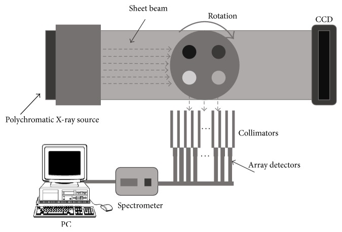
X-ray fluorescence computed tomography (XFCT) based on sheet beam can save a huge amount of time to obtain a whole set of projections using synchrotron. However, it is clearly unpractical for most biomedical research laboratories. In this paper, polychromatic X-ray fluorescence computed tomography with sheet-beam geometry is tested by Monte Carlo simulation. First, two phantoms (A and B) filled with PMMA are used to simulate imaging process through GEANT 4. Phantom A contains several GNP-loaded regions with the same size (10 mm) in height and diameter but different Au weight concentration ranging from 0.3% to 1.8%. Phantom B contains twelve GNP-loaded regions with the same Au weight concentration (1.6%) but different diameter ranging from 1 mm to 9 mm. Second, discretized presentation of imaging model is established to reconstruct more accurate XFCT images. Third, XFCT images of phantoms A and B are reconstructed by filter back-projection (FBP) and maximum likelihood expectation maximization (MLEM) with and without correction, respectively. Contrast-to-noise ratio (CNR) is calculated to evaluate all the reconstructed images. Our results show that it is feasible for sheet-beam XFCT system based on polychromatic X-ray source and the discretized imaging model can be used to reconstruct more accurate images.
期刊介绍:
The International Journal of Biomedical Imaging is managed by a board of editors comprising internationally renowned active researchers. The journal is freely accessible online and also offered for purchase in print format. It employs a web-based review system to ensure swift turnaround times while maintaining high standards. In addition to regular issues, special issues are organized by guest editors. The subject areas covered include (but are not limited to):
Digital radiography and tomosynthesis
X-ray computed tomography (CT)
Magnetic resonance imaging (MRI)
Single photon emission computed tomography (SPECT)
Positron emission tomography (PET)
Ultrasound imaging
Diffuse optical tomography, coherence, fluorescence, bioluminescence tomography, impedance tomography
Neutron imaging for biomedical applications
Magnetic and optical spectroscopy, and optical biopsy
Optical, electron, scanning tunneling/atomic force microscopy
Small animal imaging
Functional, cellular, and molecular imaging
Imaging assays for screening and molecular analysis
Microarray image analysis and bioinformatics
Emerging biomedical imaging techniques
Imaging modality fusion
Biomedical imaging instrumentation
Biomedical image processing, pattern recognition, and analysis
Biomedical image visualization, compression, transmission, and storage
Imaging and modeling related to systems biology and systems biomedicine
Applied mathematics, applied physics, and chemistry related to biomedical imaging
Grid-enabling technology for biomedical imaging and informatics

 求助内容:
求助内容: 应助结果提醒方式:


