Jane S Kim, Richard L Scawn, Bradford W Lee, Jonathan H Lin, Bobby S Korn, Don O Kikkawa
{"title":"假面性眼眶结节病伴孤立性眼外肌受累。","authors":"Jane S Kim, Richard L Scawn, Bradford W Lee, Jonathan H Lin, Bobby S Korn, Don O Kikkawa","doi":"10.2174/1874364101610010140","DOIUrl":null,"url":null,"abstract":"<p><p>Two patients, previously diagnosed and treated for euthyroid, autoantibody-negative thyroid eye disease, presented with active orbitopathy. An atypical disease course and presentation prompted orbital biopsy. Extraocular muscle histopathology demonstrated noncaseating granulomatous inflammation consistent with presumed orbital sarcoidosis involving multiple extraocular muscles, including the inferior oblique in one of the cases. These two cases emphasize the importance of a broad differential diagnosis and the utility of an orbital biopsy in the context of an unusual disease presentation or poor treatment response. The patients' clinical course is discussed alongside important clinical signs, imaging findings, and biopsy results that support a diagnosis of isolated orbital sarcoidosis.</p>","PeriodicalId":512318,"journal":{"name":"The Open Ophthalmology Journal","volume":"10 ","pages":"140-145"},"PeriodicalIF":0.0000,"publicationDate":"2016-04-29","publicationTypes":"Journal Article","fieldsOfStudy":null,"isOpenAccess":false,"openAccessPdf":"https://ftp.ncbi.nlm.nih.gov/pub/pmc/oa_pdf/89/7e/TOOPHTJ-10-140.PMC5396127.pdf","citationCount":"9","resultStr":"{\"title\":\"Masquerading Orbital Sarcoidosis with Isolated Extraocular Muscle Involvement.\",\"authors\":\"Jane S Kim, Richard L Scawn, Bradford W Lee, Jonathan H Lin, Bobby S Korn, Don O Kikkawa\",\"doi\":\"10.2174/1874364101610010140\",\"DOIUrl\":null,\"url\":null,\"abstract\":\"<p><p>Two patients, previously diagnosed and treated for euthyroid, autoantibody-negative thyroid eye disease, presented with active orbitopathy. An atypical disease course and presentation prompted orbital biopsy. Extraocular muscle histopathology demonstrated noncaseating granulomatous inflammation consistent with presumed orbital sarcoidosis involving multiple extraocular muscles, including the inferior oblique in one of the cases. These two cases emphasize the importance of a broad differential diagnosis and the utility of an orbital biopsy in the context of an unusual disease presentation or poor treatment response. The patients' clinical course is discussed alongside important clinical signs, imaging findings, and biopsy results that support a diagnosis of isolated orbital sarcoidosis.</p>\",\"PeriodicalId\":512318,\"journal\":{\"name\":\"The Open Ophthalmology Journal\",\"volume\":\"10 \",\"pages\":\"140-145\"},\"PeriodicalIF\":0.0000,\"publicationDate\":\"2016-04-29\",\"publicationTypes\":\"Journal Article\",\"fieldsOfStudy\":null,\"isOpenAccess\":false,\"openAccessPdf\":\"https://ftp.ncbi.nlm.nih.gov/pub/pmc/oa_pdf/89/7e/TOOPHTJ-10-140.PMC5396127.pdf\",\"citationCount\":\"9\",\"resultStr\":null,\"platform\":\"Semanticscholar\",\"paperid\":null,\"PeriodicalName\":\"The Open Ophthalmology Journal\",\"FirstCategoryId\":\"1085\",\"ListUrlMain\":\"https://doi.org/10.2174/1874364101610010140\",\"RegionNum\":0,\"RegionCategory\":null,\"ArticlePicture\":[],\"TitleCN\":null,\"AbstractTextCN\":null,\"PMCID\":null,\"EPubDate\":\"2016/1/1 0:00:00\",\"PubModel\":\"eCollection\",\"JCR\":\"\",\"JCRName\":\"\",\"Score\":null,\"Total\":0}","platform":"Semanticscholar","paperid":null,"PeriodicalName":"The Open Ophthalmology Journal","FirstCategoryId":"1085","ListUrlMain":"https://doi.org/10.2174/1874364101610010140","RegionNum":0,"RegionCategory":null,"ArticlePicture":[],"TitleCN":null,"AbstractTextCN":null,"PMCID":null,"EPubDate":"2016/1/1 0:00:00","PubModel":"eCollection","JCR":"","JCRName":"","Score":null,"Total":0}
Masquerading Orbital Sarcoidosis with Isolated Extraocular Muscle Involvement.
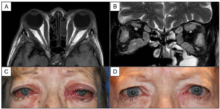
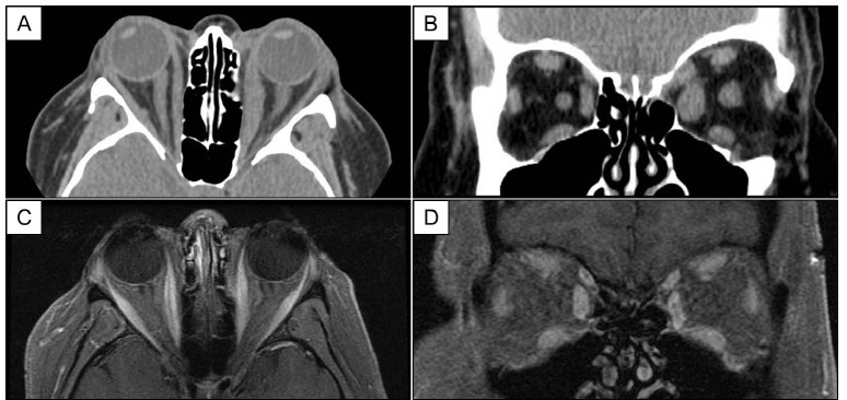
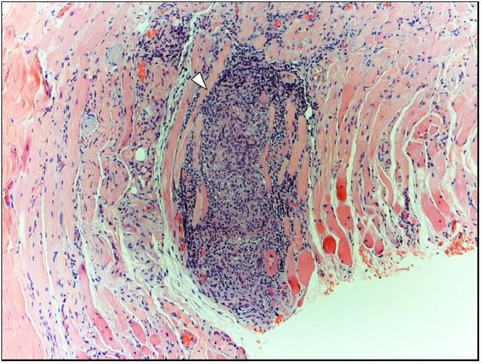
Two patients, previously diagnosed and treated for euthyroid, autoantibody-negative thyroid eye disease, presented with active orbitopathy. An atypical disease course and presentation prompted orbital biopsy. Extraocular muscle histopathology demonstrated noncaseating granulomatous inflammation consistent with presumed orbital sarcoidosis involving multiple extraocular muscles, including the inferior oblique in one of the cases. These two cases emphasize the importance of a broad differential diagnosis and the utility of an orbital biopsy in the context of an unusual disease presentation or poor treatment response. The patients' clinical course is discussed alongside important clinical signs, imaging findings, and biopsy results that support a diagnosis of isolated orbital sarcoidosis.

 求助内容:
求助内容: 应助结果提醒方式:


