{"title":"牙结石 修复龋齿。","authors":"Paul H Keyes, Thomas E Rams","doi":"10.13188/2377-987x.1000017","DOIUrl":null,"url":null,"abstract":"<p><strong>Background: </strong>An inverse relationship between dental calculus mineralization and dental caries demineralization on teeth has been noted in some studies. Dental calculus may even form superficial layers over existing dental caries and arrest their progression, but this phenomenon has been only rarely documented and infrequently considered in the field of Cariology. To further assess the occurrence of dental calculus arrest of dental caries, this study evaluated a large number of extracted human teeth for the presence and location of dental caries, dental calculus, and dental plaque biofilms.</p><p><strong>Materials and methods: </strong>A total of 1,200 teeth were preserved in 10% buffered formal saline, and viewed while moist by a single experienced examiner using a research stereomicroscope at 15-25× magnification. Representative teeth were sectioned and photographed, and their dental plaque biofilms subjected to gram-stain examination with light microscopy at 100× magnification.</p><p><strong>Results: </strong>Dental calculus was observed on 1,140 (95%) of the extracted human teeth, and no dental carious lesions were found underlying dental calculus-covered surfaces on 1,139 of these teeth. However, dental calculus arrest of dental caries was found on one (0.54%) of 187 evaluated teeth that presented with unrestored proximal enamel caries. On the distal surface of a maxillary premolar tooth, dental calculus mineralization filled the outer surface cavitation of an incipient dental caries lesion. The dental calculus-covered carious lesion extended only slightly into enamel, and exhibited a brown pigmentation characteristic of inactive or arrested dental caries. In contrast, the tooth's mesial surface, without a superficial layer of dental calculus, had a large carious lesion going through enamel and deep into dentin.</p><p><strong>Conclusions: </strong>These observations further document the potential protective effects of dental calculus mineralization against dental caries.</p>","PeriodicalId":91029,"journal":{"name":"Journal of oral biology (Northborough, Mass.)","volume":"3 1","pages":""},"PeriodicalIF":0.0000,"publicationDate":"2016-01-01","publicationTypes":"Journal Article","fieldsOfStudy":null,"isOpenAccess":false,"openAccessPdf":"https://www.ncbi.nlm.nih.gov/pmc/articles/PMC4950958/pdf/","citationCount":"0","resultStr":"{\"title\":\"Dental Calculus Arrest of Dental Caries.\",\"authors\":\"Paul H Keyes, Thomas E Rams\",\"doi\":\"10.13188/2377-987x.1000017\",\"DOIUrl\":null,\"url\":null,\"abstract\":\"<p><strong>Background: </strong>An inverse relationship between dental calculus mineralization and dental caries demineralization on teeth has been noted in some studies. Dental calculus may even form superficial layers over existing dental caries and arrest their progression, but this phenomenon has been only rarely documented and infrequently considered in the field of Cariology. To further assess the occurrence of dental calculus arrest of dental caries, this study evaluated a large number of extracted human teeth for the presence and location of dental caries, dental calculus, and dental plaque biofilms.</p><p><strong>Materials and methods: </strong>A total of 1,200 teeth were preserved in 10% buffered formal saline, and viewed while moist by a single experienced examiner using a research stereomicroscope at 15-25× magnification. Representative teeth were sectioned and photographed, and their dental plaque biofilms subjected to gram-stain examination with light microscopy at 100× magnification.</p><p><strong>Results: </strong>Dental calculus was observed on 1,140 (95%) of the extracted human teeth, and no dental carious lesions were found underlying dental calculus-covered surfaces on 1,139 of these teeth. However, dental calculus arrest of dental caries was found on one (0.54%) of 187 evaluated teeth that presented with unrestored proximal enamel caries. On the distal surface of a maxillary premolar tooth, dental calculus mineralization filled the outer surface cavitation of an incipient dental caries lesion. The dental calculus-covered carious lesion extended only slightly into enamel, and exhibited a brown pigmentation characteristic of inactive or arrested dental caries. In contrast, the tooth's mesial surface, without a superficial layer of dental calculus, had a large carious lesion going through enamel and deep into dentin.</p><p><strong>Conclusions: </strong>These observations further document the potential protective effects of dental calculus mineralization against dental caries.</p>\",\"PeriodicalId\":91029,\"journal\":{\"name\":\"Journal of oral biology (Northborough, Mass.)\",\"volume\":\"3 1\",\"pages\":\"\"},\"PeriodicalIF\":0.0000,\"publicationDate\":\"2016-01-01\",\"publicationTypes\":\"Journal Article\",\"fieldsOfStudy\":null,\"isOpenAccess\":false,\"openAccessPdf\":\"https://www.ncbi.nlm.nih.gov/pmc/articles/PMC4950958/pdf/\",\"citationCount\":\"0\",\"resultStr\":null,\"platform\":\"Semanticscholar\",\"paperid\":null,\"PeriodicalName\":\"Journal of oral biology (Northborough, Mass.)\",\"FirstCategoryId\":\"1085\",\"ListUrlMain\":\"https://doi.org/10.13188/2377-987x.1000017\",\"RegionNum\":0,\"RegionCategory\":null,\"ArticlePicture\":[],\"TitleCN\":null,\"AbstractTextCN\":null,\"PMCID\":null,\"EPubDate\":\"2016/2/12 0:00:00\",\"PubModel\":\"Epub\",\"JCR\":\"\",\"JCRName\":\"\",\"Score\":null,\"Total\":0}","platform":"Semanticscholar","paperid":null,"PeriodicalName":"Journal of oral biology (Northborough, Mass.)","FirstCategoryId":"1085","ListUrlMain":"https://doi.org/10.13188/2377-987x.1000017","RegionNum":0,"RegionCategory":null,"ArticlePicture":[],"TitleCN":null,"AbstractTextCN":null,"PMCID":null,"EPubDate":"2016/2/12 0:00:00","PubModel":"Epub","JCR":"","JCRName":"","Score":null,"Total":0}
Background: An inverse relationship between dental calculus mineralization and dental caries demineralization on teeth has been noted in some studies. Dental calculus may even form superficial layers over existing dental caries and arrest their progression, but this phenomenon has been only rarely documented and infrequently considered in the field of Cariology. To further assess the occurrence of dental calculus arrest of dental caries, this study evaluated a large number of extracted human teeth for the presence and location of dental caries, dental calculus, and dental plaque biofilms.
Materials and methods: A total of 1,200 teeth were preserved in 10% buffered formal saline, and viewed while moist by a single experienced examiner using a research stereomicroscope at 15-25× magnification. Representative teeth were sectioned and photographed, and their dental plaque biofilms subjected to gram-stain examination with light microscopy at 100× magnification.
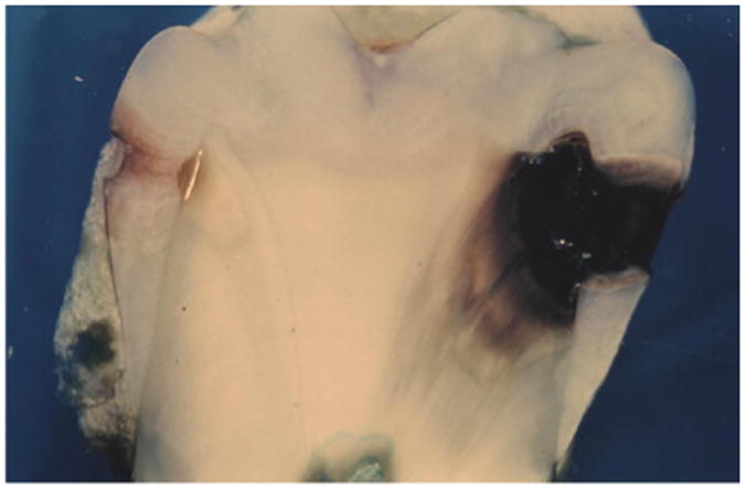
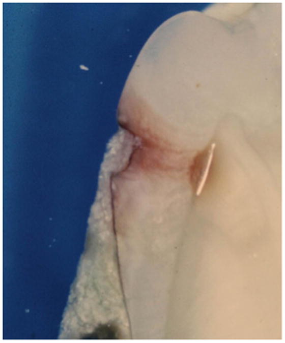
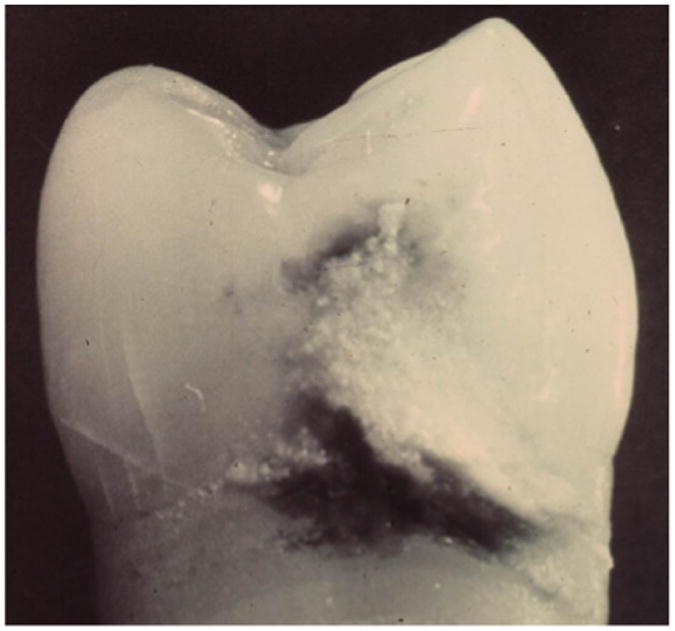
Results: Dental calculus was observed on 1,140 (95%) of the extracted human teeth, and no dental carious lesions were found underlying dental calculus-covered surfaces on 1,139 of these teeth. However, dental calculus arrest of dental caries was found on one (0.54%) of 187 evaluated teeth that presented with unrestored proximal enamel caries. On the distal surface of a maxillary premolar tooth, dental calculus mineralization filled the outer surface cavitation of an incipient dental caries lesion. The dental calculus-covered carious lesion extended only slightly into enamel, and exhibited a brown pigmentation characteristic of inactive or arrested dental caries. In contrast, the tooth's mesial surface, without a superficial layer of dental calculus, had a large carious lesion going through enamel and deep into dentin.
Conclusions: These observations further document the potential protective effects of dental calculus mineralization against dental caries.

 求助内容:
求助内容: 应助结果提醒方式:


