{"title":"人类骨骼中有限的骨小梁密度异质性。","authors":"Habiba Chirchir","doi":"10.1155/2016/9295383","DOIUrl":null,"url":null,"abstract":"<p><p>There is evidence for variation in trabecular bone density and volume within an individual skeleton, albeit in a few anatomical sites, which is partly dependent on mechanical loading. However, little is known regarding the basic variation in trabecular bone density throughout the skeleton in healthy human adults. This is because research on bone density has been confined to a few skeletal elements, which can be readily measured using available imaging technology particularly in clinical settings. This study comprehensively investigates the distribution of trabecular bone density within the human skeleton in nine skeletal sites (femur, proximal and distal tibia, third metatarsal, humerus, ulna, radius, third metacarpal, and axis) in a sample of N = 20 individuals (11 males and 9 females). pQCT results showed that the proximal ulna (mean = 231.3 mg/cm(3)) and axis vertebra (mean = 234.3 mg/cm(3)) displayed significantly greater (p < 0.01) trabecular bone density than other elements, whereas there was no significant variation among the rest of the elements (p > 0.01). The homogeneity of the majority of elements suggests that these sites are potentially responsive to site-specific genetic factors. Secondly, the lack of correlation between elements (p > 0.05) suggests that density measurements of one anatomical region are not necessarily accurate measures of other anatomical regions. </p>","PeriodicalId":89526,"journal":{"name":"Anatomy research international","volume":"2016 ","pages":"9295383"},"PeriodicalIF":0.0000,"publicationDate":"2016-01-01","publicationTypes":"Journal Article","fieldsOfStudy":null,"isOpenAccess":false,"openAccessPdf":"https://sci-hub-pdf.com/10.1155/2016/9295383","citationCount":"19","resultStr":"{\"title\":\"Limited Trabecular Bone Density Heterogeneity in the Human Skeleton.\",\"authors\":\"Habiba Chirchir\",\"doi\":\"10.1155/2016/9295383\",\"DOIUrl\":null,\"url\":null,\"abstract\":\"<p><p>There is evidence for variation in trabecular bone density and volume within an individual skeleton, albeit in a few anatomical sites, which is partly dependent on mechanical loading. However, little is known regarding the basic variation in trabecular bone density throughout the skeleton in healthy human adults. This is because research on bone density has been confined to a few skeletal elements, which can be readily measured using available imaging technology particularly in clinical settings. This study comprehensively investigates the distribution of trabecular bone density within the human skeleton in nine skeletal sites (femur, proximal and distal tibia, third metatarsal, humerus, ulna, radius, third metacarpal, and axis) in a sample of N = 20 individuals (11 males and 9 females). pQCT results showed that the proximal ulna (mean = 231.3 mg/cm(3)) and axis vertebra (mean = 234.3 mg/cm(3)) displayed significantly greater (p < 0.01) trabecular bone density than other elements, whereas there was no significant variation among the rest of the elements (p > 0.01). The homogeneity of the majority of elements suggests that these sites are potentially responsive to site-specific genetic factors. Secondly, the lack of correlation between elements (p > 0.05) suggests that density measurements of one anatomical region are not necessarily accurate measures of other anatomical regions. </p>\",\"PeriodicalId\":89526,\"journal\":{\"name\":\"Anatomy research international\",\"volume\":\"2016 \",\"pages\":\"9295383\"},\"PeriodicalIF\":0.0000,\"publicationDate\":\"2016-01-01\",\"publicationTypes\":\"Journal Article\",\"fieldsOfStudy\":null,\"isOpenAccess\":false,\"openAccessPdf\":\"https://sci-hub-pdf.com/10.1155/2016/9295383\",\"citationCount\":\"19\",\"resultStr\":null,\"platform\":\"Semanticscholar\",\"paperid\":null,\"PeriodicalName\":\"Anatomy research international\",\"FirstCategoryId\":\"1085\",\"ListUrlMain\":\"https://doi.org/10.1155/2016/9295383\",\"RegionNum\":0,\"RegionCategory\":null,\"ArticlePicture\":[],\"TitleCN\":null,\"AbstractTextCN\":null,\"PMCID\":null,\"EPubDate\":\"2016/4/11 0:00:00\",\"PubModel\":\"Epub\",\"JCR\":\"\",\"JCRName\":\"\",\"Score\":null,\"Total\":0}","platform":"Semanticscholar","paperid":null,"PeriodicalName":"Anatomy research international","FirstCategoryId":"1085","ListUrlMain":"https://doi.org/10.1155/2016/9295383","RegionNum":0,"RegionCategory":null,"ArticlePicture":[],"TitleCN":null,"AbstractTextCN":null,"PMCID":null,"EPubDate":"2016/4/11 0:00:00","PubModel":"Epub","JCR":"","JCRName":"","Score":null,"Total":0}
Limited Trabecular Bone Density Heterogeneity in the Human Skeleton.
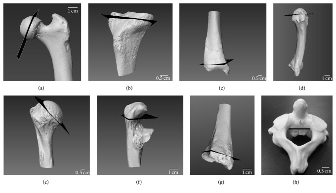
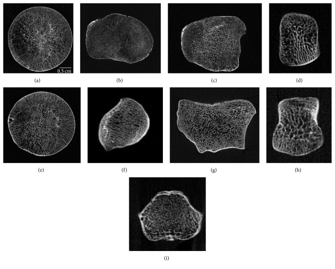
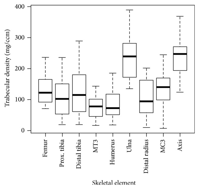
There is evidence for variation in trabecular bone density and volume within an individual skeleton, albeit in a few anatomical sites, which is partly dependent on mechanical loading. However, little is known regarding the basic variation in trabecular bone density throughout the skeleton in healthy human adults. This is because research on bone density has been confined to a few skeletal elements, which can be readily measured using available imaging technology particularly in clinical settings. This study comprehensively investigates the distribution of trabecular bone density within the human skeleton in nine skeletal sites (femur, proximal and distal tibia, third metatarsal, humerus, ulna, radius, third metacarpal, and axis) in a sample of N = 20 individuals (11 males and 9 females). pQCT results showed that the proximal ulna (mean = 231.3 mg/cm(3)) and axis vertebra (mean = 234.3 mg/cm(3)) displayed significantly greater (p < 0.01) trabecular bone density than other elements, whereas there was no significant variation among the rest of the elements (p > 0.01). The homogeneity of the majority of elements suggests that these sites are potentially responsive to site-specific genetic factors. Secondly, the lack of correlation between elements (p > 0.05) suggests that density measurements of one anatomical region are not necessarily accurate measures of other anatomical regions.

 求助内容:
求助内容: 应助结果提醒方式:


