Santosh Kaur Sangari, Paul-Michel Dossous, Thomas Heineman, Estomih Phillip Mtui
{"title":"典型颈椎横孔的尺寸和解剖变异。","authors":"Santosh Kaur Sangari, Paul-Michel Dossous, Thomas Heineman, Estomih Phillip Mtui","doi":"10.1155/2015/391823","DOIUrl":null,"url":null,"abstract":"<p><p>The study was conducted on random sample of seventy-one dried, typical cervical vertebrae (C3-C6). The data on the age, sex, and built was not available. Using vernier calipers with 0.01 mm accuracy, the anteroposterior and transverse diameters of transverse foramina and their distance from the medial margin of the uncinate process were measured bilaterally. The mean diameter of the right/left transverse foramen varied from 2.54 mm to 7.79 mm (mean = 5.55 ± 0.87 mm) and from 2.65 mm to 7.35 mm (mean = 5.48 ± 0.77 mm), respectively. The transverse foramen was less than 3.5 mm in three vertebrae on the right and two on the left. The osteocytes observed in 21.3% of specimens and the narrow transverse foramen may place patients at risk for vertebrobasilar insufficiency or thrombus formation. The mean distance of the transverse foramen from the medial margin of uncinate process is an important landmark to avoid vertebral artery laceration and was 5.0 ± 0.87 mm (range: 3.5-7.9 mm) on the right and 5.0 ± 1.0 mm (range: 3.2-7.7 mm) on the left side. No statistically significant difference was observed between the right and left sides. The accessory transverse foramina seen in 24% of vertebrae suggest duplications or fenestrations in the vertebral artery. </p>","PeriodicalId":89526,"journal":{"name":"Anatomy research international","volume":"2015 ","pages":"391823"},"PeriodicalIF":0.0000,"publicationDate":"2015-01-01","publicationTypes":"Journal Article","fieldsOfStudy":null,"isOpenAccess":false,"openAccessPdf":"https://sci-hub-pdf.com/10.1155/2015/391823","citationCount":"26","resultStr":"{\"title\":\"Dimensions and Anatomical Variants of the Foramen Transversarium of Typical Cervical Vertebrae.\",\"authors\":\"Santosh Kaur Sangari, Paul-Michel Dossous, Thomas Heineman, Estomih Phillip Mtui\",\"doi\":\"10.1155/2015/391823\",\"DOIUrl\":null,\"url\":null,\"abstract\":\"<p><p>The study was conducted on random sample of seventy-one dried, typical cervical vertebrae (C3-C6). The data on the age, sex, and built was not available. Using vernier calipers with 0.01 mm accuracy, the anteroposterior and transverse diameters of transverse foramina and their distance from the medial margin of the uncinate process were measured bilaterally. The mean diameter of the right/left transverse foramen varied from 2.54 mm to 7.79 mm (mean = 5.55 ± 0.87 mm) and from 2.65 mm to 7.35 mm (mean = 5.48 ± 0.77 mm), respectively. The transverse foramen was less than 3.5 mm in three vertebrae on the right and two on the left. The osteocytes observed in 21.3% of specimens and the narrow transverse foramen may place patients at risk for vertebrobasilar insufficiency or thrombus formation. The mean distance of the transverse foramen from the medial margin of uncinate process is an important landmark to avoid vertebral artery laceration and was 5.0 ± 0.87 mm (range: 3.5-7.9 mm) on the right and 5.0 ± 1.0 mm (range: 3.2-7.7 mm) on the left side. No statistically significant difference was observed between the right and left sides. The accessory transverse foramina seen in 24% of vertebrae suggest duplications or fenestrations in the vertebral artery. </p>\",\"PeriodicalId\":89526,\"journal\":{\"name\":\"Anatomy research international\",\"volume\":\"2015 \",\"pages\":\"391823\"},\"PeriodicalIF\":0.0000,\"publicationDate\":\"2015-01-01\",\"publicationTypes\":\"Journal Article\",\"fieldsOfStudy\":null,\"isOpenAccess\":false,\"openAccessPdf\":\"https://sci-hub-pdf.com/10.1155/2015/391823\",\"citationCount\":\"26\",\"resultStr\":null,\"platform\":\"Semanticscholar\",\"paperid\":null,\"PeriodicalName\":\"Anatomy research international\",\"FirstCategoryId\":\"1085\",\"ListUrlMain\":\"https://doi.org/10.1155/2015/391823\",\"RegionNum\":0,\"RegionCategory\":null,\"ArticlePicture\":[],\"TitleCN\":null,\"AbstractTextCN\":null,\"PMCID\":null,\"EPubDate\":\"2015/9/10 0:00:00\",\"PubModel\":\"Epub\",\"JCR\":\"\",\"JCRName\":\"\",\"Score\":null,\"Total\":0}","platform":"Semanticscholar","paperid":null,"PeriodicalName":"Anatomy research international","FirstCategoryId":"1085","ListUrlMain":"https://doi.org/10.1155/2015/391823","RegionNum":0,"RegionCategory":null,"ArticlePicture":[],"TitleCN":null,"AbstractTextCN":null,"PMCID":null,"EPubDate":"2015/9/10 0:00:00","PubModel":"Epub","JCR":"","JCRName":"","Score":null,"Total":0}
Dimensions and Anatomical Variants of the Foramen Transversarium of Typical Cervical Vertebrae.
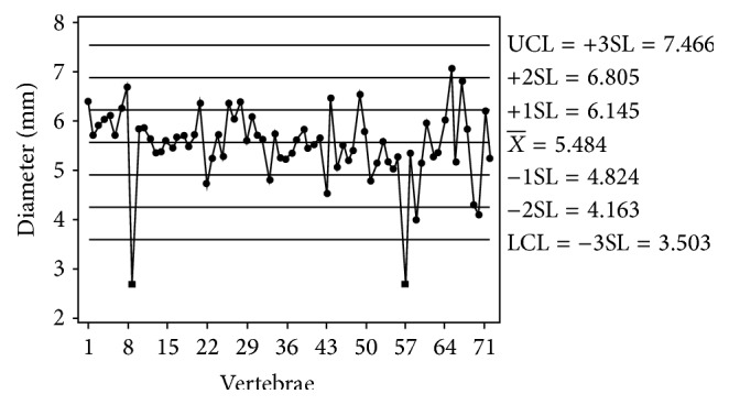
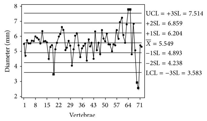
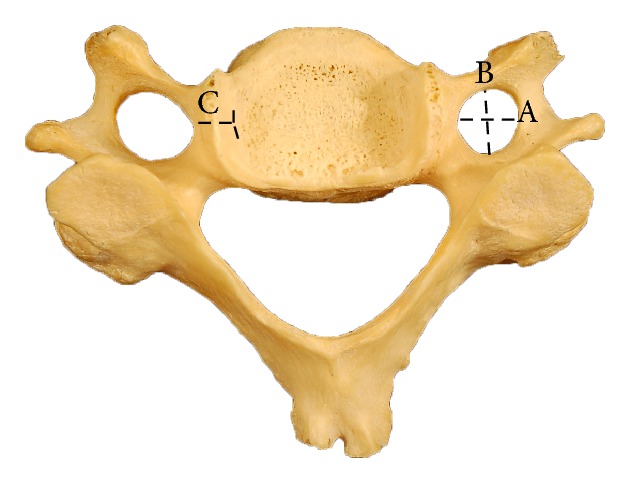
The study was conducted on random sample of seventy-one dried, typical cervical vertebrae (C3-C6). The data on the age, sex, and built was not available. Using vernier calipers with 0.01 mm accuracy, the anteroposterior and transverse diameters of transverse foramina and their distance from the medial margin of the uncinate process were measured bilaterally. The mean diameter of the right/left transverse foramen varied from 2.54 mm to 7.79 mm (mean = 5.55 ± 0.87 mm) and from 2.65 mm to 7.35 mm (mean = 5.48 ± 0.77 mm), respectively. The transverse foramen was less than 3.5 mm in three vertebrae on the right and two on the left. The osteocytes observed in 21.3% of specimens and the narrow transverse foramen may place patients at risk for vertebrobasilar insufficiency or thrombus formation. The mean distance of the transverse foramen from the medial margin of uncinate process is an important landmark to avoid vertebral artery laceration and was 5.0 ± 0.87 mm (range: 3.5-7.9 mm) on the right and 5.0 ± 1.0 mm (range: 3.2-7.7 mm) on the left side. No statistically significant difference was observed between the right and left sides. The accessory transverse foramina seen in 24% of vertebrae suggest duplications or fenestrations in the vertebral artery.

 求助内容:
求助内容: 应助结果提醒方式:


