S Rochelle Lewis, Siobhan P Ellison, John J Dascanio, David S Lindsay, Robert M Gogal, Stephen R Werre, Naveen Surendran, Meghan E Breen, Bettina M Heid, Frank M Andrews, Virginia A Buechner-Maxwell, Sharon G Witonsky
{"title":"实验性神经元肌囊虫感染对马模型免疫的影响。","authors":"S Rochelle Lewis, Siobhan P Ellison, John J Dascanio, David S Lindsay, Robert M Gogal, Stephen R Werre, Naveen Surendran, Meghan E Breen, Bettina M Heid, Frank M Andrews, Virginia A Buechner-Maxwell, Sharon G Witonsky","doi":"10.1155/2014/239495","DOIUrl":null,"url":null,"abstract":"<p><p>Sarcocystis neurona is the most common cause of Equine Protozoal Myeloencephalitis (EPM), affecting 0.5-1% horses in the United States during their lifetimes. The objective of this study was to evaluate the equine immune responses in an experimentally induced Sarcocystis neurona infection model. Neurologic parameters were recorded prior to and throughout the 70-day study by blinded investigators. Recombinant SnSAG1 ELISA for serum and CSF were used to confirm and track disease progression. All experimentally infected horses displayed neurologic signs after infection. Neutrophils, monocytes, and lymphocytes from infected horses displayed significantly delayed apoptosis at some time points. Cell proliferation was significantly increased in S. neurona-infected horses when stimulated nonspecifically with PMA/I but significantly decreased when stimulated with S. neurona compared to controls. Collectively, our results suggest that horses experimentally infected with S. neurona manifest impaired antigen specific response to S. neurona, which could be a function of altered antigen presentation, lack of antigen recognition, or both. </p>","PeriodicalId":91135,"journal":{"name":"Journal of veterinary medicine","volume":"2014 ","pages":"239495"},"PeriodicalIF":0.0000,"publicationDate":"2014-01-01","publicationTypes":"Journal Article","fieldsOfStudy":null,"isOpenAccess":false,"openAccessPdf":"https://sci-hub-pdf.com/10.1155/2014/239495","citationCount":"11","resultStr":"{\"title\":\"Effects of Experimental Sarcocystis neurona-Induced Infection on Immunity in an Equine Model.\",\"authors\":\"S Rochelle Lewis, Siobhan P Ellison, John J Dascanio, David S Lindsay, Robert M Gogal, Stephen R Werre, Naveen Surendran, Meghan E Breen, Bettina M Heid, Frank M Andrews, Virginia A Buechner-Maxwell, Sharon G Witonsky\",\"doi\":\"10.1155/2014/239495\",\"DOIUrl\":null,\"url\":null,\"abstract\":\"<p><p>Sarcocystis neurona is the most common cause of Equine Protozoal Myeloencephalitis (EPM), affecting 0.5-1% horses in the United States during their lifetimes. The objective of this study was to evaluate the equine immune responses in an experimentally induced Sarcocystis neurona infection model. Neurologic parameters were recorded prior to and throughout the 70-day study by blinded investigators. Recombinant SnSAG1 ELISA for serum and CSF were used to confirm and track disease progression. All experimentally infected horses displayed neurologic signs after infection. Neutrophils, monocytes, and lymphocytes from infected horses displayed significantly delayed apoptosis at some time points. Cell proliferation was significantly increased in S. neurona-infected horses when stimulated nonspecifically with PMA/I but significantly decreased when stimulated with S. neurona compared to controls. Collectively, our results suggest that horses experimentally infected with S. neurona manifest impaired antigen specific response to S. neurona, which could be a function of altered antigen presentation, lack of antigen recognition, or both. </p>\",\"PeriodicalId\":91135,\"journal\":{\"name\":\"Journal of veterinary medicine\",\"volume\":\"2014 \",\"pages\":\"239495\"},\"PeriodicalIF\":0.0000,\"publicationDate\":\"2014-01-01\",\"publicationTypes\":\"Journal Article\",\"fieldsOfStudy\":null,\"isOpenAccess\":false,\"openAccessPdf\":\"https://sci-hub-pdf.com/10.1155/2014/239495\",\"citationCount\":\"11\",\"resultStr\":null,\"platform\":\"Semanticscholar\",\"paperid\":null,\"PeriodicalName\":\"Journal of veterinary medicine\",\"FirstCategoryId\":\"1085\",\"ListUrlMain\":\"https://doi.org/10.1155/2014/239495\",\"RegionNum\":0,\"RegionCategory\":null,\"ArticlePicture\":[],\"TitleCN\":null,\"AbstractTextCN\":null,\"PMCID\":null,\"EPubDate\":\"2014/11/12 0:00:00\",\"PubModel\":\"Epub\",\"JCR\":\"\",\"JCRName\":\"\",\"Score\":null,\"Total\":0}","platform":"Semanticscholar","paperid":null,"PeriodicalName":"Journal of veterinary medicine","FirstCategoryId":"1085","ListUrlMain":"https://doi.org/10.1155/2014/239495","RegionNum":0,"RegionCategory":null,"ArticlePicture":[],"TitleCN":null,"AbstractTextCN":null,"PMCID":null,"EPubDate":"2014/11/12 0:00:00","PubModel":"Epub","JCR":"","JCRName":"","Score":null,"Total":0}
Effects of Experimental Sarcocystis neurona-Induced Infection on Immunity in an Equine Model.
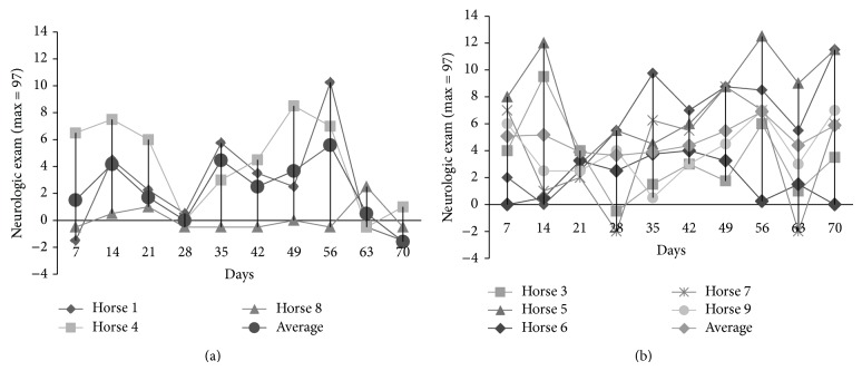
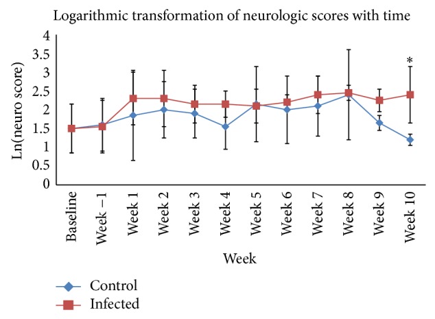
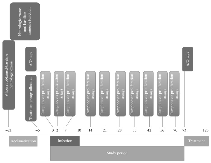
Sarcocystis neurona is the most common cause of Equine Protozoal Myeloencephalitis (EPM), affecting 0.5-1% horses in the United States during their lifetimes. The objective of this study was to evaluate the equine immune responses in an experimentally induced Sarcocystis neurona infection model. Neurologic parameters were recorded prior to and throughout the 70-day study by blinded investigators. Recombinant SnSAG1 ELISA for serum and CSF were used to confirm and track disease progression. All experimentally infected horses displayed neurologic signs after infection. Neutrophils, monocytes, and lymphocytes from infected horses displayed significantly delayed apoptosis at some time points. Cell proliferation was significantly increased in S. neurona-infected horses when stimulated nonspecifically with PMA/I but significantly decreased when stimulated with S. neurona compared to controls. Collectively, our results suggest that horses experimentally infected with S. neurona manifest impaired antigen specific response to S. neurona, which could be a function of altered antigen presentation, lack of antigen recognition, or both.

 求助内容:
求助内容: 应助结果提醒方式:


