{"title":"斯里兰卡人群下颌第一恒磨牙根形和根管形态的变异。","authors":"Roshan Peiris, Uthpala Malwatte, Janak Abayakoon, Anuradha Wettasinghe","doi":"10.1155/2015/803671","DOIUrl":null,"url":null,"abstract":"<p><p>The present study was conducted to determine the number of roots and morphology of the root canal system of permanent mandibular first molars (M1) in a Sri Lankan population. Sample of 529 M1 teeth was used. The number of roots was examined and the lengths of the mesial and distal roots were measured to the nearest 0.01 mm. Vacuum injection protocol was used to inject China ink into the root canal system, making it transparent. Root canal morphology was recorded using Vertucci's classification. Presence of furcation canals, position of lateral canals, intercanal communications, level of bifurcation, and convergence of the root canal system were recorded. M1 showed three roots in 4.1% of the sample. Commonest root canal morphology of the mesial root was type IV and the distal root was type I. The level of bifurcation of the root canals was commonly observed in the cervical one-third of the root while convergence was observed in the apical one-third in both roots. Prevalence of three rooted mandibular first molars is less than 5%. Mesial root showed the most variable canal morphology. Prevalence of furcation canals was 1.5% while that of middle mesial canals was 0.2%. </p>","PeriodicalId":89526,"journal":{"name":"Anatomy research international","volume":"2015 ","pages":"803671"},"PeriodicalIF":0.0000,"publicationDate":"2015-01-01","publicationTypes":"Journal Article","fieldsOfStudy":null,"isOpenAccess":false,"openAccessPdf":"https://sci-hub-pdf.com/10.1155/2015/803671","citationCount":"9","resultStr":"{\"title\":\"Variations in the Root Form and Root Canal Morphology of Permanent Mandibular First Molars in a Sri Lankan Population.\",\"authors\":\"Roshan Peiris, Uthpala Malwatte, Janak Abayakoon, Anuradha Wettasinghe\",\"doi\":\"10.1155/2015/803671\",\"DOIUrl\":null,\"url\":null,\"abstract\":\"<p><p>The present study was conducted to determine the number of roots and morphology of the root canal system of permanent mandibular first molars (M1) in a Sri Lankan population. Sample of 529 M1 teeth was used. The number of roots was examined and the lengths of the mesial and distal roots were measured to the nearest 0.01 mm. Vacuum injection protocol was used to inject China ink into the root canal system, making it transparent. Root canal morphology was recorded using Vertucci's classification. Presence of furcation canals, position of lateral canals, intercanal communications, level of bifurcation, and convergence of the root canal system were recorded. M1 showed three roots in 4.1% of the sample. Commonest root canal morphology of the mesial root was type IV and the distal root was type I. The level of bifurcation of the root canals was commonly observed in the cervical one-third of the root while convergence was observed in the apical one-third in both roots. Prevalence of three rooted mandibular first molars is less than 5%. Mesial root showed the most variable canal morphology. Prevalence of furcation canals was 1.5% while that of middle mesial canals was 0.2%. </p>\",\"PeriodicalId\":89526,\"journal\":{\"name\":\"Anatomy research international\",\"volume\":\"2015 \",\"pages\":\"803671\"},\"PeriodicalIF\":0.0000,\"publicationDate\":\"2015-01-01\",\"publicationTypes\":\"Journal Article\",\"fieldsOfStudy\":null,\"isOpenAccess\":false,\"openAccessPdf\":\"https://sci-hub-pdf.com/10.1155/2015/803671\",\"citationCount\":\"9\",\"resultStr\":null,\"platform\":\"Semanticscholar\",\"paperid\":null,\"PeriodicalName\":\"Anatomy research international\",\"FirstCategoryId\":\"1085\",\"ListUrlMain\":\"https://doi.org/10.1155/2015/803671\",\"RegionNum\":0,\"RegionCategory\":null,\"ArticlePicture\":[],\"TitleCN\":null,\"AbstractTextCN\":null,\"PMCID\":null,\"EPubDate\":\"2015/8/13 0:00:00\",\"PubModel\":\"Epub\",\"JCR\":\"\",\"JCRName\":\"\",\"Score\":null,\"Total\":0}","platform":"Semanticscholar","paperid":null,"PeriodicalName":"Anatomy research international","FirstCategoryId":"1085","ListUrlMain":"https://doi.org/10.1155/2015/803671","RegionNum":0,"RegionCategory":null,"ArticlePicture":[],"TitleCN":null,"AbstractTextCN":null,"PMCID":null,"EPubDate":"2015/8/13 0:00:00","PubModel":"Epub","JCR":"","JCRName":"","Score":null,"Total":0}
Variations in the Root Form and Root Canal Morphology of Permanent Mandibular First Molars in a Sri Lankan Population.
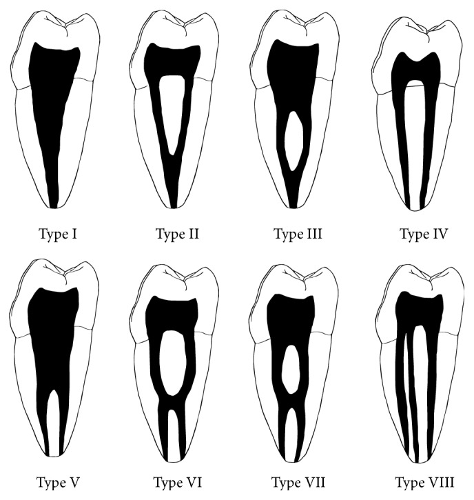
The present study was conducted to determine the number of roots and morphology of the root canal system of permanent mandibular first molars (M1) in a Sri Lankan population. Sample of 529 M1 teeth was used. The number of roots was examined and the lengths of the mesial and distal roots were measured to the nearest 0.01 mm. Vacuum injection protocol was used to inject China ink into the root canal system, making it transparent. Root canal morphology was recorded using Vertucci's classification. Presence of furcation canals, position of lateral canals, intercanal communications, level of bifurcation, and convergence of the root canal system were recorded. M1 showed three roots in 4.1% of the sample. Commonest root canal morphology of the mesial root was type IV and the distal root was type I. The level of bifurcation of the root canals was commonly observed in the cervical one-third of the root while convergence was observed in the apical one-third in both roots. Prevalence of three rooted mandibular first molars is less than 5%. Mesial root showed the most variable canal morphology. Prevalence of furcation canals was 1.5% while that of middle mesial canals was 0.2%.


 求助内容:
求助内容: 应助结果提醒方式:


