Aarti Sarwal, Yash Patel, Ralph D'Agostino, Patrick Brown, Stacey Q Wolfe, Cheryl Bushnell, Casey Glass, Pamela Duncan
{"title":"评价护理点颅脑超声检测脑出血可行性的探索性研究。","authors":"Aarti Sarwal, Yash Patel, Ralph D'Agostino, Patrick Brown, Stacey Q Wolfe, Cheryl Bushnell, Casey Glass, Pamela Duncan","doi":"10.1186/s13089-022-00289-z","DOIUrl":null,"url":null,"abstract":"<p><strong>Background: </strong>Limited studies have evaluated the use of ultrasound for detection of intracerebral hemorrhage (ICH) using diagnostic ultrasound Transcranial Doppler machines in adults. The feasibility of ICH detection using Point of care Ultrasound (POCUS) machines has not been explored. We evaluated the feasibility of using cranial POCUS B mode imaging performed using intensive care unit (ICU) POCUS device for ICH detection with a secondary goal of mapping optimal imaging technique and brain topography likely to affect sensitivity and specificity of ICH detection with POCUS.</p><p><strong>Materials and methods: </strong>After obtaining IRB approval, a blinded investigator performed cranial ultrasound (Fujifilm, Sonosite<sup>®</sup> Xporte, transcranial and abdominal presets) through temporal windows on 11 patients with intracerebral pathology within 72 h of last CT/MRI (computed tomography scan/magnetic resonance imaging) brain after being admitted to a neurocritical care unit in Aug 2020 and Nov 2020-Mar 2021. Images were then compared to patient's CT/MRI to inform topography. Inferential statistics were reported.</p><p><strong>Results: </strong>Mean age was 57 (28-77 years) and 6/11 were female. Six patients were diagnosed with ICH, 3 with ischemic stroke, 1 subarachnoid hemorrhage, and 1 brain tumor. The sensitivity and specificity of point of care diagnosis of ICH compared to CT/MRI brain was 100% and 50%, respectively. Mean time between ultrasound scan and CT/MRI was 13.3 h (21 min-39 h). Falx cerebri, choroid calcification and midbrain-related artifacts were the most reproducible hyperechoic signals. Abdominal preset on high gain yielded less artifact than Transcranial Doppler preset for cranial B mode imaging. False positive ICH diagnosis was attributed to intracerebral tumor and midbrain-related artifact.</p><p><strong>Conclusions: </strong>Our exploratory analysis yielded preliminary data on use of point of care cranial ultrasound for ICH diagnosis to inform imaging techniques, cranial topography on B mode and sample size estimation for future studies to evaluate sensitivity and specificity of cranial POCUS in adult patients. This pilot study is limited by small sample size and over representation of ICH in the study. Cranial POCUS is feasible using POCUS machines and may have potential as a screening tool if validated in adequately powered studies.</p>","PeriodicalId":36911,"journal":{"name":"Ultrasound Journal","volume":" ","pages":"40"},"PeriodicalIF":3.4000,"publicationDate":"2022-10-17","publicationTypes":"Journal Article","fieldsOfStudy":null,"isOpenAccess":false,"openAccessPdf":"https://www.ncbi.nlm.nih.gov/pmc/articles/PMC9576831/pdf/","citationCount":"4","resultStr":"{\"title\":\"Exploratory study to assess feasibility of intracerebral hemorrhage detection by point of care cranial ultrasound.\",\"authors\":\"Aarti Sarwal, Yash Patel, Ralph D'Agostino, Patrick Brown, Stacey Q Wolfe, Cheryl Bushnell, Casey Glass, Pamela Duncan\",\"doi\":\"10.1186/s13089-022-00289-z\",\"DOIUrl\":null,\"url\":null,\"abstract\":\"<p><strong>Background: </strong>Limited studies have evaluated the use of ultrasound for detection of intracerebral hemorrhage (ICH) using diagnostic ultrasound Transcranial Doppler machines in adults. The feasibility of ICH detection using Point of care Ultrasound (POCUS) machines has not been explored. We evaluated the feasibility of using cranial POCUS B mode imaging performed using intensive care unit (ICU) POCUS device for ICH detection with a secondary goal of mapping optimal imaging technique and brain topography likely to affect sensitivity and specificity of ICH detection with POCUS.</p><p><strong>Materials and methods: </strong>After obtaining IRB approval, a blinded investigator performed cranial ultrasound (Fujifilm, Sonosite<sup>®</sup> Xporte, transcranial and abdominal presets) through temporal windows on 11 patients with intracerebral pathology within 72 h of last CT/MRI (computed tomography scan/magnetic resonance imaging) brain after being admitted to a neurocritical care unit in Aug 2020 and Nov 2020-Mar 2021. Images were then compared to patient's CT/MRI to inform topography. Inferential statistics were reported.</p><p><strong>Results: </strong>Mean age was 57 (28-77 years) and 6/11 were female. Six patients were diagnosed with ICH, 3 with ischemic stroke, 1 subarachnoid hemorrhage, and 1 brain tumor. The sensitivity and specificity of point of care diagnosis of ICH compared to CT/MRI brain was 100% and 50%, respectively. Mean time between ultrasound scan and CT/MRI was 13.3 h (21 min-39 h). Falx cerebri, choroid calcification and midbrain-related artifacts were the most reproducible hyperechoic signals. Abdominal preset on high gain yielded less artifact than Transcranial Doppler preset for cranial B mode imaging. False positive ICH diagnosis was attributed to intracerebral tumor and midbrain-related artifact.</p><p><strong>Conclusions: </strong>Our exploratory analysis yielded preliminary data on use of point of care cranial ultrasound for ICH diagnosis to inform imaging techniques, cranial topography on B mode and sample size estimation for future studies to evaluate sensitivity and specificity of cranial POCUS in adult patients. This pilot study is limited by small sample size and over representation of ICH in the study. Cranial POCUS is feasible using POCUS machines and may have potential as a screening tool if validated in adequately powered studies.</p>\",\"PeriodicalId\":36911,\"journal\":{\"name\":\"Ultrasound Journal\",\"volume\":\" \",\"pages\":\"40\"},\"PeriodicalIF\":3.4000,\"publicationDate\":\"2022-10-17\",\"publicationTypes\":\"Journal Article\",\"fieldsOfStudy\":null,\"isOpenAccess\":false,\"openAccessPdf\":\"https://www.ncbi.nlm.nih.gov/pmc/articles/PMC9576831/pdf/\",\"citationCount\":\"4\",\"resultStr\":null,\"platform\":\"Semanticscholar\",\"paperid\":null,\"PeriodicalName\":\"Ultrasound Journal\",\"FirstCategoryId\":\"1085\",\"ListUrlMain\":\"https://doi.org/10.1186/s13089-022-00289-z\",\"RegionNum\":0,\"RegionCategory\":null,\"ArticlePicture\":[],\"TitleCN\":null,\"AbstractTextCN\":null,\"PMCID\":null,\"EPubDate\":\"\",\"PubModel\":\"\",\"JCR\":\"Q2\",\"JCRName\":\"Medicine\",\"Score\":null,\"Total\":0}","platform":"Semanticscholar","paperid":null,"PeriodicalName":"Ultrasound Journal","FirstCategoryId":"1085","ListUrlMain":"https://doi.org/10.1186/s13089-022-00289-z","RegionNum":0,"RegionCategory":null,"ArticlePicture":[],"TitleCN":null,"AbstractTextCN":null,"PMCID":null,"EPubDate":"","PubModel":"","JCR":"Q2","JCRName":"Medicine","Score":null,"Total":0}
引用次数: 4
摘要
背景:有限的研究评估了使用经颅多普勒超声诊断成人脑出血(ICH)的超声检测。使用点护理超声(POCUS)机器检测脑出血的可行性尚未探讨。我们评估了使用重症监护病房(ICU) POCUS设备进行颅内POCUS B模式成像用于脑出血检测的可行性,其次要目标是绘制最佳成像技术和可能影响POCUS脑出血检测敏感性和特异性的脑地形。材料和方法:在获得IRB批准后,一名盲法研究者在2020年8月和2020年11月至2021年3月入住神经危重症监护病房后的最后一次CT/MRI(计算机断层扫描/磁共振成像)72小时内,通过时间窗对11名脑内病理患者进行了颅超声(Fujifilm, Sonosite®Xporte,经颅和腹部预设)。然后将图像与患者的CT/MRI进行比较,以了解地形。进行了推论统计。结果:平均年龄57岁(28 ~ 77岁),女性6/11。诊断为脑出血6例,缺血性脑卒中3例,蛛网膜下腔出血1例,脑肿瘤1例。与CT/MRI脑相比较,护理点诊断脑出血的敏感性和特异性分别为100%和50%。超声与CT/MRI扫描的平均间隔时间为13.3 h (21 min-39 h)。大脑镰、脉膜钙化和中脑相关伪影是重现性最强的高回声信号。腹部高增益预设比经颅多普勒预设产生更少的伪影。脑出血假阳性诊断归因于颅内肿瘤和中脑相关伪影。结论:我们的探索性分析获得了使用护理点颅超声诊断脑出血的初步数据,为未来研究评估成人患者颅POCUS的敏感性和特异性提供了成像技术、B模式颅形貌和样本量估计的信息。这项初步研究受到样本量小和研究中ICH的过度代表性的限制。颅POCUS是可行的使用POCUS机器,可能有潜力作为筛选工具,如果在充分有力的研究验证。
Exploratory study to assess feasibility of intracerebral hemorrhage detection by point of care cranial ultrasound.
Background: Limited studies have evaluated the use of ultrasound for detection of intracerebral hemorrhage (ICH) using diagnostic ultrasound Transcranial Doppler machines in adults. The feasibility of ICH detection using Point of care Ultrasound (POCUS) machines has not been explored. We evaluated the feasibility of using cranial POCUS B mode imaging performed using intensive care unit (ICU) POCUS device for ICH detection with a secondary goal of mapping optimal imaging technique and brain topography likely to affect sensitivity and specificity of ICH detection with POCUS.
Materials and methods: After obtaining IRB approval, a blinded investigator performed cranial ultrasound (Fujifilm, Sonosite® Xporte, transcranial and abdominal presets) through temporal windows on 11 patients with intracerebral pathology within 72 h of last CT/MRI (computed tomography scan/magnetic resonance imaging) brain after being admitted to a neurocritical care unit in Aug 2020 and Nov 2020-Mar 2021. Images were then compared to patient's CT/MRI to inform topography. Inferential statistics were reported.
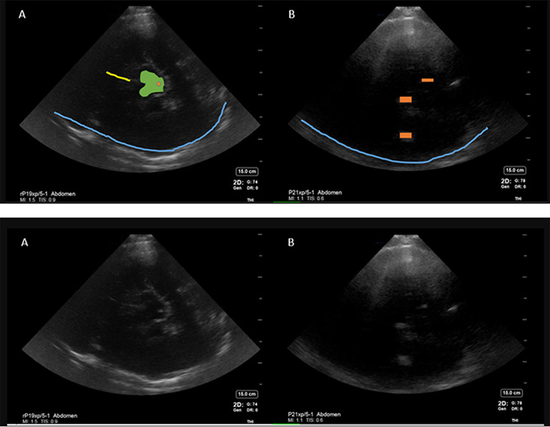
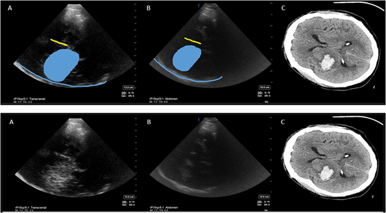
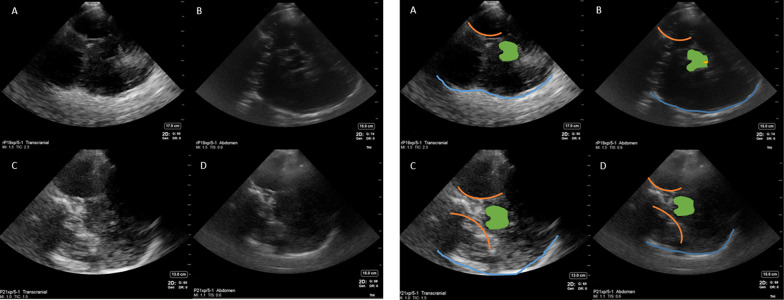
Results: Mean age was 57 (28-77 years) and 6/11 were female. Six patients were diagnosed with ICH, 3 with ischemic stroke, 1 subarachnoid hemorrhage, and 1 brain tumor. The sensitivity and specificity of point of care diagnosis of ICH compared to CT/MRI brain was 100% and 50%, respectively. Mean time between ultrasound scan and CT/MRI was 13.3 h (21 min-39 h). Falx cerebri, choroid calcification and midbrain-related artifacts were the most reproducible hyperechoic signals. Abdominal preset on high gain yielded less artifact than Transcranial Doppler preset for cranial B mode imaging. False positive ICH diagnosis was attributed to intracerebral tumor and midbrain-related artifact.
Conclusions: Our exploratory analysis yielded preliminary data on use of point of care cranial ultrasound for ICH diagnosis to inform imaging techniques, cranial topography on B mode and sample size estimation for future studies to evaluate sensitivity and specificity of cranial POCUS in adult patients. This pilot study is limited by small sample size and over representation of ICH in the study. Cranial POCUS is feasible using POCUS machines and may have potential as a screening tool if validated in adequately powered studies.

 求助内容:
求助内容: 应助结果提醒方式:


