Ozlem Ozkale Yavuz, Ercan Ayaz, Yılmaz Yıldız, Ayca Akgoz Karaosmanoglu, Elif Bulut, H Serap Kalkanoglu Sivri, Kader K Oguz
{"title":"粘多糖病VI型患儿眼壁厚度增加,眼球体积减小。","authors":"Ozlem Ozkale Yavuz, Ercan Ayaz, Yılmaz Yıldız, Ayca Akgoz Karaosmanoglu, Elif Bulut, H Serap Kalkanoglu Sivri, Kader K Oguz","doi":"10.5152/dir.2022.21372","DOIUrl":null,"url":null,"abstract":"<p><p>PURPOSE lthough clinical ophthalmologic findings have been reported, no study documented magnetic resonance imaging (MRI) findings in mucopolysaccharidosis (MPS) type VI. The aim of this study was to determine the ophthalmologic imaging findings of MPS type VI in the pediatric age group retrospectively. METHODS Brain MRIs of 10 patients with MPS type VI and 49 healthy children were evaluated independently by two pediatric radiologists for the following characteristics: globe volume, ocular wall thickness, and optic nerve sheath diameter for each orbit. The means of the measurement of each group were compared by using an independent t-test. Agreement and bias between reviewers were assessed by intra-class correlation coefficients (ICC). RESULTS A total of 59 children [32 girls (54.23%), 27 boys (45.77%); age range, 4-16 years; mean age, 10.37 ± 3.73 years] were included in the study. Statistical analysis revealed smaller eyeballs and thicker ocular walls of patients with MPS type VI (P < .001 and P < .001, respectively). However, there was no statistically significant difference in terms of optic nerve sheath diameter between the two groups (P=.648). CONCLUSION Patients with MPS type VI displayed reduced globe volumes and increased ocular wall thicknesses compared to the healthy children. Therefore, we recommend that ophthalmologic imaging findings might prove to be an auxiliary tool in the diagnosis of MPS patients.</p>","PeriodicalId":50582,"journal":{"name":"Diagnostic and Interventional Radiology","volume":" ","pages":"516-521"},"PeriodicalIF":1.7000,"publicationDate":"2022-09-01","publicationTypes":"Journal Article","fieldsOfStudy":null,"isOpenAccess":false,"openAccessPdf":"https://ftp.ncbi.nlm.nih.gov/pub/pmc/oa_pdf/48/62/dir-28-5-516.PMC9682595.pdf","citationCount":"0","resultStr":"{\"title\":\"Increased ocular wall thickness and decreased globe volume in children with mucopolysaccharidosis type VI.\",\"authors\":\"Ozlem Ozkale Yavuz, Ercan Ayaz, Yılmaz Yıldız, Ayca Akgoz Karaosmanoglu, Elif Bulut, H Serap Kalkanoglu Sivri, Kader K Oguz\",\"doi\":\"10.5152/dir.2022.21372\",\"DOIUrl\":null,\"url\":null,\"abstract\":\"<p><p>PURPOSE lthough clinical ophthalmologic findings have been reported, no study documented magnetic resonance imaging (MRI) findings in mucopolysaccharidosis (MPS) type VI. The aim of this study was to determine the ophthalmologic imaging findings of MPS type VI in the pediatric age group retrospectively. METHODS Brain MRIs of 10 patients with MPS type VI and 49 healthy children were evaluated independently by two pediatric radiologists for the following characteristics: globe volume, ocular wall thickness, and optic nerve sheath diameter for each orbit. The means of the measurement of each group were compared by using an independent t-test. Agreement and bias between reviewers were assessed by intra-class correlation coefficients (ICC). RESULTS A total of 59 children [32 girls (54.23%), 27 boys (45.77%); age range, 4-16 years; mean age, 10.37 ± 3.73 years] were included in the study. Statistical analysis revealed smaller eyeballs and thicker ocular walls of patients with MPS type VI (P < .001 and P < .001, respectively). However, there was no statistically significant difference in terms of optic nerve sheath diameter between the two groups (P=.648). CONCLUSION Patients with MPS type VI displayed reduced globe volumes and increased ocular wall thicknesses compared to the healthy children. Therefore, we recommend that ophthalmologic imaging findings might prove to be an auxiliary tool in the diagnosis of MPS patients.</p>\",\"PeriodicalId\":50582,\"journal\":{\"name\":\"Diagnostic and Interventional Radiology\",\"volume\":\" \",\"pages\":\"516-521\"},\"PeriodicalIF\":1.7000,\"publicationDate\":\"2022-09-01\",\"publicationTypes\":\"Journal Article\",\"fieldsOfStudy\":null,\"isOpenAccess\":false,\"openAccessPdf\":\"https://ftp.ncbi.nlm.nih.gov/pub/pmc/oa_pdf/48/62/dir-28-5-516.PMC9682595.pdf\",\"citationCount\":\"0\",\"resultStr\":null,\"platform\":\"Semanticscholar\",\"paperid\":null,\"PeriodicalName\":\"Diagnostic and Interventional Radiology\",\"FirstCategoryId\":\"3\",\"ListUrlMain\":\"https://doi.org/10.5152/dir.2022.21372\",\"RegionNum\":4,\"RegionCategory\":\"医学\",\"ArticlePicture\":[],\"TitleCN\":null,\"AbstractTextCN\":null,\"PMCID\":null,\"EPubDate\":\"\",\"PubModel\":\"\",\"JCR\":\"Q2\",\"JCRName\":\"Medicine\",\"Score\":null,\"Total\":0}","platform":"Semanticscholar","paperid":null,"PeriodicalName":"Diagnostic and Interventional Radiology","FirstCategoryId":"3","ListUrlMain":"https://doi.org/10.5152/dir.2022.21372","RegionNum":4,"RegionCategory":"医学","ArticlePicture":[],"TitleCN":null,"AbstractTextCN":null,"PMCID":null,"EPubDate":"","PubModel":"","JCR":"Q2","JCRName":"Medicine","Score":null,"Total":0}
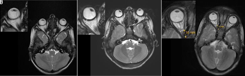
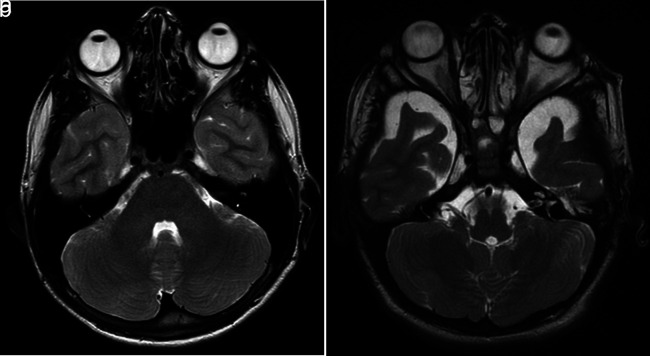
Increased ocular wall thickness and decreased globe volume in children with mucopolysaccharidosis type VI.
PURPOSE lthough clinical ophthalmologic findings have been reported, no study documented magnetic resonance imaging (MRI) findings in mucopolysaccharidosis (MPS) type VI. The aim of this study was to determine the ophthalmologic imaging findings of MPS type VI in the pediatric age group retrospectively. METHODS Brain MRIs of 10 patients with MPS type VI and 49 healthy children were evaluated independently by two pediatric radiologists for the following characteristics: globe volume, ocular wall thickness, and optic nerve sheath diameter for each orbit. The means of the measurement of each group were compared by using an independent t-test. Agreement and bias between reviewers were assessed by intra-class correlation coefficients (ICC). RESULTS A total of 59 children [32 girls (54.23%), 27 boys (45.77%); age range, 4-16 years; mean age, 10.37 ± 3.73 years] were included in the study. Statistical analysis revealed smaller eyeballs and thicker ocular walls of patients with MPS type VI (P < .001 and P < .001, respectively). However, there was no statistically significant difference in terms of optic nerve sheath diameter between the two groups (P=.648). CONCLUSION Patients with MPS type VI displayed reduced globe volumes and increased ocular wall thicknesses compared to the healthy children. Therefore, we recommend that ophthalmologic imaging findings might prove to be an auxiliary tool in the diagnosis of MPS patients.
期刊介绍:
Diagnostic and Interventional Radiology (Diagn Interv Radiol) is the open access, online-only official publication of Turkish Society of Radiology. It is published bimonthly and the journal’s publication language is English.
The journal is a medium for original articles, reviews, pictorial essays, technical notes related to all fields of diagnostic and interventional radiology.


 求助内容:
求助内容: 应助结果提醒方式:


