Lang Wang, Mimi Jia, Xiaoling Wen, Jiang Shen, Hanfeng Yang
{"title":"磁共振成像特征对肝细胞癌微血管浸润的诊断价值:荟萃分析。","authors":"Lang Wang, Mimi Jia, Xiaoling Wen, Jiang Shen, Hanfeng Yang","doi":"10.5152/dir.2022.21108","DOIUrl":null,"url":null,"abstract":"<p><p>PURPOSE This systematic review and meta-analysis of conventional enhanced magnetic resonance imaging (MRI) were conducted to evaluate the diagnostic performance of imaging features of microvascular invasion (MVI) prediction in hepatocellular carcinoma (HCC). METHODS Relevant studies on diagnosing MVI in HCC by MRI were searched in the MEDLINE, PUBMED, EMBASE, Cochrane library, and Web of Science databases. The pooled mean sensitivity and specificity were calculated using a random effects model. The corresponding positive likelihood ratio (PLR), negative likelihood ratio (NLR), and pooled diagnostic odds ratio (DOR) were calculated. The summary receiver operating characteristic (SROC) curve was used to summarize the overall diagnostic accuracy. Diagnostic performance was evaluated by determining the area under the curve (AUC). Regression analysis by subgroup and sensitivity analysis were used to explore potential sources of heterogeneity. RESULTS A total of 19 studies comprising 1920 HCC patients with 2033 tumors were ultimately enrolled. For the signs of the presence of peritumoral enhancement in the arterial phase, peritumoral hypointensity in the hepatobiliary phase, irregular non-smooth margin, and rim-like enhancement in the arterial phase, the pooled sensitivity values, the pooled specificity values, the pooled PLR values, the pooled NLR values, the pooled DOR values, and the values of the AUC of SROC curves were determined. CONCLUSION The conventional MRI features for predicting MVI showed poor diagnostic performance in HCC. Only signs of the presence of peritumoral enhancement in the arterial phase showed a moderate diagnostic accuracy.</p>","PeriodicalId":50582,"journal":{"name":"Diagnostic and Interventional Radiology","volume":" ","pages":"428-440"},"PeriodicalIF":1.7000,"publicationDate":"2022-09-01","publicationTypes":"Journal Article","fieldsOfStudy":null,"isOpenAccess":false,"openAccessPdf":"https://ftp.ncbi.nlm.nih.gov/pub/pmc/oa_pdf/7c/92/dir-28-5-428.PMC9682567.pdf","citationCount":"0","resultStr":"{\"title\":\"Diagnostic value of magnetic resonance imaging features of microvascular invasion in hepatocellular carcinoma: a meta-analysis.\",\"authors\":\"Lang Wang, Mimi Jia, Xiaoling Wen, Jiang Shen, Hanfeng Yang\",\"doi\":\"10.5152/dir.2022.21108\",\"DOIUrl\":null,\"url\":null,\"abstract\":\"<p><p>PURPOSE This systematic review and meta-analysis of conventional enhanced magnetic resonance imaging (MRI) were conducted to evaluate the diagnostic performance of imaging features of microvascular invasion (MVI) prediction in hepatocellular carcinoma (HCC). METHODS Relevant studies on diagnosing MVI in HCC by MRI were searched in the MEDLINE, PUBMED, EMBASE, Cochrane library, and Web of Science databases. The pooled mean sensitivity and specificity were calculated using a random effects model. The corresponding positive likelihood ratio (PLR), negative likelihood ratio (NLR), and pooled diagnostic odds ratio (DOR) were calculated. The summary receiver operating characteristic (SROC) curve was used to summarize the overall diagnostic accuracy. Diagnostic performance was evaluated by determining the area under the curve (AUC). Regression analysis by subgroup and sensitivity analysis were used to explore potential sources of heterogeneity. RESULTS A total of 19 studies comprising 1920 HCC patients with 2033 tumors were ultimately enrolled. For the signs of the presence of peritumoral enhancement in the arterial phase, peritumoral hypointensity in the hepatobiliary phase, irregular non-smooth margin, and rim-like enhancement in the arterial phase, the pooled sensitivity values, the pooled specificity values, the pooled PLR values, the pooled NLR values, the pooled DOR values, and the values of the AUC of SROC curves were determined. CONCLUSION The conventional MRI features for predicting MVI showed poor diagnostic performance in HCC. Only signs of the presence of peritumoral enhancement in the arterial phase showed a moderate diagnostic accuracy.</p>\",\"PeriodicalId\":50582,\"journal\":{\"name\":\"Diagnostic and Interventional Radiology\",\"volume\":\" \",\"pages\":\"428-440\"},\"PeriodicalIF\":1.7000,\"publicationDate\":\"2022-09-01\",\"publicationTypes\":\"Journal Article\",\"fieldsOfStudy\":null,\"isOpenAccess\":false,\"openAccessPdf\":\"https://ftp.ncbi.nlm.nih.gov/pub/pmc/oa_pdf/7c/92/dir-28-5-428.PMC9682567.pdf\",\"citationCount\":\"0\",\"resultStr\":null,\"platform\":\"Semanticscholar\",\"paperid\":null,\"PeriodicalName\":\"Diagnostic and Interventional Radiology\",\"FirstCategoryId\":\"3\",\"ListUrlMain\":\"https://doi.org/10.5152/dir.2022.21108\",\"RegionNum\":4,\"RegionCategory\":\"医学\",\"ArticlePicture\":[],\"TitleCN\":null,\"AbstractTextCN\":null,\"PMCID\":null,\"EPubDate\":\"\",\"PubModel\":\"\",\"JCR\":\"Q2\",\"JCRName\":\"Medicine\",\"Score\":null,\"Total\":0}","platform":"Semanticscholar","paperid":null,"PeriodicalName":"Diagnostic and Interventional Radiology","FirstCategoryId":"3","ListUrlMain":"https://doi.org/10.5152/dir.2022.21108","RegionNum":4,"RegionCategory":"医学","ArticlePicture":[],"TitleCN":null,"AbstractTextCN":null,"PMCID":null,"EPubDate":"","PubModel":"","JCR":"Q2","JCRName":"Medicine","Score":null,"Total":0}
引用次数: 0
摘要
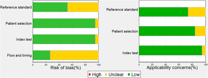
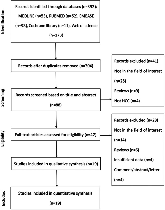
目的:本研究对常规增强磁共振成像(MRI)进行系统回顾和荟萃分析,以评估成像特征对肝细胞癌(HCC)微血管侵袭(MVI)预测的诊断价值。方法检索MEDLINE、PUBMED、EMBASE、Cochrane图书馆和Web of Science数据库中MRI诊断肝癌MVI的相关研究。采用随机效应模型计算合并平均敏感性和特异性。计算相应的阳性似然比(PLR)、阴性似然比(NLR)和合并诊断优势比(DOR)。采用总受者工作特征(SROC)曲线总结总体诊断准确率。通过测定曲线下面积(AUC)来评估诊断效果。采用亚组回归分析和敏感性分析探讨异质性的潜在来源。结果共纳入19项研究,包括1920例HCC患者和2033个肿瘤。对于动脉期肿瘤周围强化、肝胆期肿瘤周围低密度、边缘不规则非光滑、动脉期边缘样强化等征象,确定SROC曲线的敏感性、特异性、PLR、NLR、DOR和AUC的综合值。结论预测MVI的常规MRI特征对HCC的诊断效果较差。只有在动脉期出现肿瘤周围强化的征象显示了中等的诊断准确性。
Diagnostic value of magnetic resonance imaging features of microvascular invasion in hepatocellular carcinoma: a meta-analysis.
PURPOSE This systematic review and meta-analysis of conventional enhanced magnetic resonance imaging (MRI) were conducted to evaluate the diagnostic performance of imaging features of microvascular invasion (MVI) prediction in hepatocellular carcinoma (HCC). METHODS Relevant studies on diagnosing MVI in HCC by MRI were searched in the MEDLINE, PUBMED, EMBASE, Cochrane library, and Web of Science databases. The pooled mean sensitivity and specificity were calculated using a random effects model. The corresponding positive likelihood ratio (PLR), negative likelihood ratio (NLR), and pooled diagnostic odds ratio (DOR) were calculated. The summary receiver operating characteristic (SROC) curve was used to summarize the overall diagnostic accuracy. Diagnostic performance was evaluated by determining the area under the curve (AUC). Regression analysis by subgroup and sensitivity analysis were used to explore potential sources of heterogeneity. RESULTS A total of 19 studies comprising 1920 HCC patients with 2033 tumors were ultimately enrolled. For the signs of the presence of peritumoral enhancement in the arterial phase, peritumoral hypointensity in the hepatobiliary phase, irregular non-smooth margin, and rim-like enhancement in the arterial phase, the pooled sensitivity values, the pooled specificity values, the pooled PLR values, the pooled NLR values, the pooled DOR values, and the values of the AUC of SROC curves were determined. CONCLUSION The conventional MRI features for predicting MVI showed poor diagnostic performance in HCC. Only signs of the presence of peritumoral enhancement in the arterial phase showed a moderate diagnostic accuracy.
期刊介绍:
Diagnostic and Interventional Radiology (Diagn Interv Radiol) is the open access, online-only official publication of Turkish Society of Radiology. It is published bimonthly and the journal’s publication language is English.
The journal is a medium for original articles, reviews, pictorial essays, technical notes related to all fields of diagnostic and interventional radiology.


 求助内容:
求助内容: 应助结果提醒方式:


