{"title":"小梁切除术和联合法氏囊-小梁切除术后早期黄斑厚度的变化","authors":"Naveed Nilforushan, Shima Loni, Parya Abdolalizadeh, Arezoo Miraftabi, Mohammad Banifatemi, Reza Rakhshan, Samira Jafari, Navid Abolfathzadeh","doi":"10.4103/joco.joco_333_21","DOIUrl":null,"url":null,"abstract":"<p><strong>Purpose: </strong>To assess postoperative changes in central retinal thickness (RT) following trabeculectomy and combined phaco-trabeculectomy using spectral domain-optical coherence tomography.</p><p><strong>Methods: </strong>In a prospective interventional comparative study, 64 consecutive glaucoma patients who underwent trabeculectomy (32 eyes) or phaco-trabeculectomy (32 eyes) were included. A macular thickness map using the Early Treatment Diabetic Retinopathy Study circles of 1 mm, 3 mm, and 6 mm was the standard to evaluate the 9-subfield thickness preoperatively and again at 1 and 3 months after surgery. Four subfields in each of the 3 mm and 6 mm rings were considered parafoveal and perifoveal regions, respectively.</p><p><strong>Results: </strong>Preoperative measurements were similar in the two groups, except patients in the combined group which were older (<i>P</i> = 0.002). The mean RT in the combined phaco-trabeculectomy group at month 1 was significantly higher than baseline measurements at central subfield retinal thickness (CSRT) (<i>P</i> = 0.01), temporal (<i>P</i> = 0.001), and inferior (<i>P</i> = 0.04) parafoveal and temporal (<i>P</i> = 0.01), superior (<i>P</i> = 0.02), and nasal (<i>P</i> < 0.001) perifoveal quadrants; however, RT changes in the trabeculectomy-only group were not statistically significant at months 1 and 3 (<i>P</i> > 0.05). The increase in the temporal perifoveal RT of the combined phaco-trabeculectomy group persisted at month 3 (<i>P</i> = 0.01), while the RT in other sectors returned to preoperative values. The two treatment groups did not differ in terms of changes in the CSRT over time (<i>P</i> = 0.37). In addition, no difference was observed between the treatment groups regarding the parafoveal RTs at each time points (0.06 ≤ <i>P</i> ≤ 0.29).</p><p><strong>Conclusions: </strong>There was no significant difference in the pattern of changes of CSRT and parafoveal RT between trabeculectomy and combined phaco-trabeculectomy treatment groups up to 3 months after surgery. Some detectable increase in RT in the combined phaco-trabeculectomy will reverse to baseline values 3 months after surgery, except in the temporal perifoveal region.</p>","PeriodicalId":15423,"journal":{"name":"Journal of Current Ophthalmology","volume":"34 2","pages":"160-166"},"PeriodicalIF":1.2000,"publicationDate":"2022-07-26","publicationTypes":"Journal Article","fieldsOfStudy":null,"isOpenAccess":false,"openAccessPdf":"https://ftp.ncbi.nlm.nih.gov/pub/pmc/oa_pdf/3e/99/JCO-34-160.PMC9487009.pdf","citationCount":"0","resultStr":"{\"title\":\"Early Macular Thickness Changes after Trabeculectomy and Combined Phaco-Trabeculectomy.\",\"authors\":\"Naveed Nilforushan, Shima Loni, Parya Abdolalizadeh, Arezoo Miraftabi, Mohammad Banifatemi, Reza Rakhshan, Samira Jafari, Navid Abolfathzadeh\",\"doi\":\"10.4103/joco.joco_333_21\",\"DOIUrl\":null,\"url\":null,\"abstract\":\"<p><strong>Purpose: </strong>To assess postoperative changes in central retinal thickness (RT) following trabeculectomy and combined phaco-trabeculectomy using spectral domain-optical coherence tomography.</p><p><strong>Methods: </strong>In a prospective interventional comparative study, 64 consecutive glaucoma patients who underwent trabeculectomy (32 eyes) or phaco-trabeculectomy (32 eyes) were included. A macular thickness map using the Early Treatment Diabetic Retinopathy Study circles of 1 mm, 3 mm, and 6 mm was the standard to evaluate the 9-subfield thickness preoperatively and again at 1 and 3 months after surgery. Four subfields in each of the 3 mm and 6 mm rings were considered parafoveal and perifoveal regions, respectively.</p><p><strong>Results: </strong>Preoperative measurements were similar in the two groups, except patients in the combined group which were older (<i>P</i> = 0.002). The mean RT in the combined phaco-trabeculectomy group at month 1 was significantly higher than baseline measurements at central subfield retinal thickness (CSRT) (<i>P</i> = 0.01), temporal (<i>P</i> = 0.001), and inferior (<i>P</i> = 0.04) parafoveal and temporal (<i>P</i> = 0.01), superior (<i>P</i> = 0.02), and nasal (<i>P</i> < 0.001) perifoveal quadrants; however, RT changes in the trabeculectomy-only group were not statistically significant at months 1 and 3 (<i>P</i> > 0.05). The increase in the temporal perifoveal RT of the combined phaco-trabeculectomy group persisted at month 3 (<i>P</i> = 0.01), while the RT in other sectors returned to preoperative values. The two treatment groups did not differ in terms of changes in the CSRT over time (<i>P</i> = 0.37). In addition, no difference was observed between the treatment groups regarding the parafoveal RTs at each time points (0.06 ≤ <i>P</i> ≤ 0.29).</p><p><strong>Conclusions: </strong>There was no significant difference in the pattern of changes of CSRT and parafoveal RT between trabeculectomy and combined phaco-trabeculectomy treatment groups up to 3 months after surgery. Some detectable increase in RT in the combined phaco-trabeculectomy will reverse to baseline values 3 months after surgery, except in the temporal perifoveal region.</p>\",\"PeriodicalId\":15423,\"journal\":{\"name\":\"Journal of Current Ophthalmology\",\"volume\":\"34 2\",\"pages\":\"160-166\"},\"PeriodicalIF\":1.2000,\"publicationDate\":\"2022-07-26\",\"publicationTypes\":\"Journal Article\",\"fieldsOfStudy\":null,\"isOpenAccess\":false,\"openAccessPdf\":\"https://ftp.ncbi.nlm.nih.gov/pub/pmc/oa_pdf/3e/99/JCO-34-160.PMC9487009.pdf\",\"citationCount\":\"0\",\"resultStr\":null,\"platform\":\"Semanticscholar\",\"paperid\":null,\"PeriodicalName\":\"Journal of Current Ophthalmology\",\"FirstCategoryId\":\"1085\",\"ListUrlMain\":\"https://doi.org/10.4103/joco.joco_333_21\",\"RegionNum\":0,\"RegionCategory\":null,\"ArticlePicture\":[],\"TitleCN\":null,\"AbstractTextCN\":null,\"PMCID\":null,\"EPubDate\":\"2022/4/1 0:00:00\",\"PubModel\":\"eCollection\",\"JCR\":\"Q3\",\"JCRName\":\"OPHTHALMOLOGY\",\"Score\":null,\"Total\":0}","platform":"Semanticscholar","paperid":null,"PeriodicalName":"Journal of Current Ophthalmology","FirstCategoryId":"1085","ListUrlMain":"https://doi.org/10.4103/joco.joco_333_21","RegionNum":0,"RegionCategory":null,"ArticlePicture":[],"TitleCN":null,"AbstractTextCN":null,"PMCID":null,"EPubDate":"2022/4/1 0:00:00","PubModel":"eCollection","JCR":"Q3","JCRName":"OPHTHALMOLOGY","Score":null,"Total":0}
Early Macular Thickness Changes after Trabeculectomy and Combined Phaco-Trabeculectomy.
Purpose: To assess postoperative changes in central retinal thickness (RT) following trabeculectomy and combined phaco-trabeculectomy using spectral domain-optical coherence tomography.
Methods: In a prospective interventional comparative study, 64 consecutive glaucoma patients who underwent trabeculectomy (32 eyes) or phaco-trabeculectomy (32 eyes) were included. A macular thickness map using the Early Treatment Diabetic Retinopathy Study circles of 1 mm, 3 mm, and 6 mm was the standard to evaluate the 9-subfield thickness preoperatively and again at 1 and 3 months after surgery. Four subfields in each of the 3 mm and 6 mm rings were considered parafoveal and perifoveal regions, respectively.
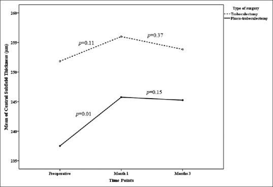
Results: Preoperative measurements were similar in the two groups, except patients in the combined group which were older (P = 0.002). The mean RT in the combined phaco-trabeculectomy group at month 1 was significantly higher than baseline measurements at central subfield retinal thickness (CSRT) (P = 0.01), temporal (P = 0.001), and inferior (P = 0.04) parafoveal and temporal (P = 0.01), superior (P = 0.02), and nasal (P < 0.001) perifoveal quadrants; however, RT changes in the trabeculectomy-only group were not statistically significant at months 1 and 3 (P > 0.05). The increase in the temporal perifoveal RT of the combined phaco-trabeculectomy group persisted at month 3 (P = 0.01), while the RT in other sectors returned to preoperative values. The two treatment groups did not differ in terms of changes in the CSRT over time (P = 0.37). In addition, no difference was observed between the treatment groups regarding the parafoveal RTs at each time points (0.06 ≤ P ≤ 0.29).
Conclusions: There was no significant difference in the pattern of changes of CSRT and parafoveal RT between trabeculectomy and combined phaco-trabeculectomy treatment groups up to 3 months after surgery. Some detectable increase in RT in the combined phaco-trabeculectomy will reverse to baseline values 3 months after surgery, except in the temporal perifoveal region.
期刊介绍:
Peer Review under the responsibility of Iranian Society of Ophthalmology Journal of Current Ophthalmology, the official publication of the Iranian Society of Ophthalmology, is a peer-reviewed, open-access, scientific journal that welcomes high quality original articles related to vision science and all fields of ophthalmology. Journal of Current Ophthalmology is the continuum of Iranian Journal of Ophthalmology published since 1969.

 求助内容:
求助内容: 应助结果提醒方式:


