{"title":"结外NK/T细胞淋巴瘤的皮肤侵犯,鼻型,各种皮肤病的临床和组织病理学模拟者。","authors":"Preeyawat Ngamdamrongkiat, Sanya Sukpanichnant, Manasmon Chairatchaneeboon, Archrob Khuhapinant, Panitta Sitthinamsuwan","doi":"10.3390/dermatopathology9030037","DOIUrl":null,"url":null,"abstract":"<p><strong>Background: </strong>Extranodal NK/T cell lymphoma, nasal type (ENK/T) with cutaneous involvement has various histopathological findings and diverse clinical manifestations.</p><p><strong>Methods: </strong>A retrospective study of cutaneous involvement of ENK/T lymphoma between 2006 and 2018 was conducted.</p><p><strong>Results: </strong>Twenty-two cases were eligible for this study. Twelve cases could be proven as secondary cutaneous involvement by ENK/T lymphoma, while the remaining could not be confirmed as primary cutaneous ENK/T lymphoma. The histopathological patterns included dermal and subcutaneous nodular infiltration pattern in 11/22 cases (50%), lobular panniculitis pattern in 6/22 cases (27.3%), interface dermatitis pattern in 4/22 cases (18.2%), and granulomatous dermatitis pattern in 1/22 case (4.5%). The median follow-up was 18.3 months. Overall, the one-year and five-year survival rates were 31.3% and 13.3%, respectively.</p><p><strong>Conclusions: </strong>A variety of histopathological patterns of cutaneous involvement by ENK/T lymphoma should be differentiated from other cutaneous lymphomas, dermatitis, and infection. When atypical medium or large-sized lymphoid cells are encountered within skin lesions, pathologists should realize these lesions can be ENK/T lymphoma, especially in cases with coexisting tumor necrosis or angioinvasion. A complete evaluation of the upper aerodigestive tract is mandatory to identify the occult primary site of ENK/T lymphoma before establishing primary cutaneous ENK/T lymphoma.</p>","PeriodicalId":42885,"journal":{"name":"Dermatopathology","volume":" ","pages":"307-320"},"PeriodicalIF":1.7000,"publicationDate":"2022-09-09","publicationTypes":"Journal Article","fieldsOfStudy":null,"isOpenAccess":false,"openAccessPdf":"https://www.ncbi.nlm.nih.gov/pmc/articles/PMC9497790/pdf/","citationCount":"1","resultStr":"{\"title\":\"Cutaneous Involvement of Extranodal NK/T Cell Lymphoma, Nasal Type, a Clinical and Histopathological Mimicker of Various Skin Diseases.\",\"authors\":\"Preeyawat Ngamdamrongkiat, Sanya Sukpanichnant, Manasmon Chairatchaneeboon, Archrob Khuhapinant, Panitta Sitthinamsuwan\",\"doi\":\"10.3390/dermatopathology9030037\",\"DOIUrl\":null,\"url\":null,\"abstract\":\"<p><strong>Background: </strong>Extranodal NK/T cell lymphoma, nasal type (ENK/T) with cutaneous involvement has various histopathological findings and diverse clinical manifestations.</p><p><strong>Methods: </strong>A retrospective study of cutaneous involvement of ENK/T lymphoma between 2006 and 2018 was conducted.</p><p><strong>Results: </strong>Twenty-two cases were eligible for this study. Twelve cases could be proven as secondary cutaneous involvement by ENK/T lymphoma, while the remaining could not be confirmed as primary cutaneous ENK/T lymphoma. The histopathological patterns included dermal and subcutaneous nodular infiltration pattern in 11/22 cases (50%), lobular panniculitis pattern in 6/22 cases (27.3%), interface dermatitis pattern in 4/22 cases (18.2%), and granulomatous dermatitis pattern in 1/22 case (4.5%). The median follow-up was 18.3 months. Overall, the one-year and five-year survival rates were 31.3% and 13.3%, respectively.</p><p><strong>Conclusions: </strong>A variety of histopathological patterns of cutaneous involvement by ENK/T lymphoma should be differentiated from other cutaneous lymphomas, dermatitis, and infection. When atypical medium or large-sized lymphoid cells are encountered within skin lesions, pathologists should realize these lesions can be ENK/T lymphoma, especially in cases with coexisting tumor necrosis or angioinvasion. A complete evaluation of the upper aerodigestive tract is mandatory to identify the occult primary site of ENK/T lymphoma before establishing primary cutaneous ENK/T lymphoma.</p>\",\"PeriodicalId\":42885,\"journal\":{\"name\":\"Dermatopathology\",\"volume\":\" \",\"pages\":\"307-320\"},\"PeriodicalIF\":1.7000,\"publicationDate\":\"2022-09-09\",\"publicationTypes\":\"Journal Article\",\"fieldsOfStudy\":null,\"isOpenAccess\":false,\"openAccessPdf\":\"https://www.ncbi.nlm.nih.gov/pmc/articles/PMC9497790/pdf/\",\"citationCount\":\"1\",\"resultStr\":null,\"platform\":\"Semanticscholar\",\"paperid\":null,\"PeriodicalName\":\"Dermatopathology\",\"FirstCategoryId\":\"1085\",\"ListUrlMain\":\"https://doi.org/10.3390/dermatopathology9030037\",\"RegionNum\":0,\"RegionCategory\":null,\"ArticlePicture\":[],\"TitleCN\":null,\"AbstractTextCN\":null,\"PMCID\":null,\"EPubDate\":\"\",\"PubModel\":\"\",\"JCR\":\"Q3\",\"JCRName\":\"DERMATOLOGY\",\"Score\":null,\"Total\":0}","platform":"Semanticscholar","paperid":null,"PeriodicalName":"Dermatopathology","FirstCategoryId":"1085","ListUrlMain":"https://doi.org/10.3390/dermatopathology9030037","RegionNum":0,"RegionCategory":null,"ArticlePicture":[],"TitleCN":null,"AbstractTextCN":null,"PMCID":null,"EPubDate":"","PubModel":"","JCR":"Q3","JCRName":"DERMATOLOGY","Score":null,"Total":0}
Cutaneous Involvement of Extranodal NK/T Cell Lymphoma, Nasal Type, a Clinical and Histopathological Mimicker of Various Skin Diseases.
Background: Extranodal NK/T cell lymphoma, nasal type (ENK/T) with cutaneous involvement has various histopathological findings and diverse clinical manifestations.
Methods: A retrospective study of cutaneous involvement of ENK/T lymphoma between 2006 and 2018 was conducted.
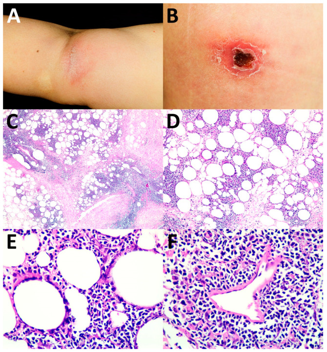
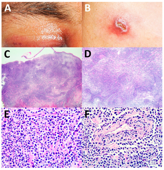
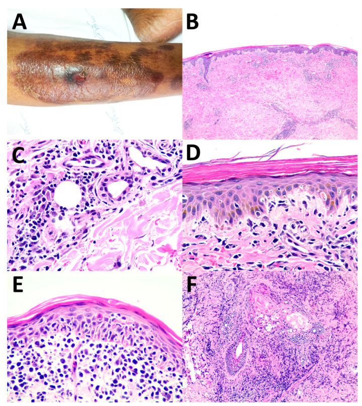
Results: Twenty-two cases were eligible for this study. Twelve cases could be proven as secondary cutaneous involvement by ENK/T lymphoma, while the remaining could not be confirmed as primary cutaneous ENK/T lymphoma. The histopathological patterns included dermal and subcutaneous nodular infiltration pattern in 11/22 cases (50%), lobular panniculitis pattern in 6/22 cases (27.3%), interface dermatitis pattern in 4/22 cases (18.2%), and granulomatous dermatitis pattern in 1/22 case (4.5%). The median follow-up was 18.3 months. Overall, the one-year and five-year survival rates were 31.3% and 13.3%, respectively.
Conclusions: A variety of histopathological patterns of cutaneous involvement by ENK/T lymphoma should be differentiated from other cutaneous lymphomas, dermatitis, and infection. When atypical medium or large-sized lymphoid cells are encountered within skin lesions, pathologists should realize these lesions can be ENK/T lymphoma, especially in cases with coexisting tumor necrosis or angioinvasion. A complete evaluation of the upper aerodigestive tract is mandatory to identify the occult primary site of ENK/T lymphoma before establishing primary cutaneous ENK/T lymphoma.

 求助内容:
求助内容: 应助结果提醒方式:


