Dingsheng Han, Yanru Zhou, Lan Zhang, Jiajia Zhang
{"title":"对比增强3D-STIR-VISTA在臂丛磁共振成像中的应用。","authors":"Dingsheng Han, Yanru Zhou, Lan Zhang, Jiajia Zhang","doi":"10.5334/jbsr.2803","DOIUrl":null,"url":null,"abstract":"<p><strong>Objective: </strong>To introduce contrast-enhanced 3D-STIR-VISTA sequence that would improve the image quality for the brachial plexus imaging and enhance the contrast between the brachial plexus and surrounding tissues.</p><p><strong>Methods: </strong>Thirty subjects (average age, 47.33 ± 15.15 years; 22 males and 8 females) were enrolled, including 7 patients with brachial plexus injuries, 4 patients with schwannomas, 1 patient with neurofibroma, 1 patient with thoracic outlet syndrome, 1 patient with metastasis, 1 patient with brachial plexus neuritis, and 15 patients without abnormal findings. Scores of unenhanced and contrast-enhanced 3D-STIR-VISTA images using a 5-point scale were compared by Wilcoxon's signed-rank test. The signal intensity (SI), signal to noise ratio (SNR), contrast to noise ratio (CNR) and contrast ratio (CR) between 3D-STIR-VISTA images without and with contrast agent were compared by the paired Student t-test.</p><p><strong>Results: </strong>The SNRs of the brachial plexus between 3D-STIR-VISTA without and with contrast agent were not significantly different, while SNRs of surrounding tissues were significantly decreased with contrast agent. The CNRs of 3D-STIR-VISTA images with contrast agent were significantly higher than that without contrast agent. The 3D-STIR-VISTA sequence with contrast agent exhibited a statistically higher CR than that without contrast agent. The average score for 3D-STIR-VISTA images with contrast agent was significantly higher than that without contrast agent.</p><p><strong>Conclusion: </strong>The 3D-STIR-VISTA sequence with contrast agent is qualitatively and quantitatively superior to that without a contrast agent. The contrast-enhanced 3D-STIR-VISTA sequence can provide distinct visualization of the brachial plexus and enhance the contrast between the brachial plexus and surrounding tissues.</p>","PeriodicalId":56282,"journal":{"name":"Journal of the Belgian Society of Radiology","volume":"106 1","pages":"75"},"PeriodicalIF":1.3000,"publicationDate":"2022-09-05","publicationTypes":"Journal Article","fieldsOfStudy":null,"isOpenAccess":false,"openAccessPdf":"https://www.ncbi.nlm.nih.gov/pmc/articles/PMC9461682/pdf/","citationCount":"0","resultStr":"{\"title\":\"The Application of Contrast-Enhanced 3D-STIR-VISTA MR Imaging of the Brachial Plexus.\",\"authors\":\"Dingsheng Han, Yanru Zhou, Lan Zhang, Jiajia Zhang\",\"doi\":\"10.5334/jbsr.2803\",\"DOIUrl\":null,\"url\":null,\"abstract\":\"<p><strong>Objective: </strong>To introduce contrast-enhanced 3D-STIR-VISTA sequence that would improve the image quality for the brachial plexus imaging and enhance the contrast between the brachial plexus and surrounding tissues.</p><p><strong>Methods: </strong>Thirty subjects (average age, 47.33 ± 15.15 years; 22 males and 8 females) were enrolled, including 7 patients with brachial plexus injuries, 4 patients with schwannomas, 1 patient with neurofibroma, 1 patient with thoracic outlet syndrome, 1 patient with metastasis, 1 patient with brachial plexus neuritis, and 15 patients without abnormal findings. Scores of unenhanced and contrast-enhanced 3D-STIR-VISTA images using a 5-point scale were compared by Wilcoxon's signed-rank test. The signal intensity (SI), signal to noise ratio (SNR), contrast to noise ratio (CNR) and contrast ratio (CR) between 3D-STIR-VISTA images without and with contrast agent were compared by the paired Student t-test.</p><p><strong>Results: </strong>The SNRs of the brachial plexus between 3D-STIR-VISTA without and with contrast agent were not significantly different, while SNRs of surrounding tissues were significantly decreased with contrast agent. The CNRs of 3D-STIR-VISTA images with contrast agent were significantly higher than that without contrast agent. The 3D-STIR-VISTA sequence with contrast agent exhibited a statistically higher CR than that without contrast agent. The average score for 3D-STIR-VISTA images with contrast agent was significantly higher than that without contrast agent.</p><p><strong>Conclusion: </strong>The 3D-STIR-VISTA sequence with contrast agent is qualitatively and quantitatively superior to that without a contrast agent. The contrast-enhanced 3D-STIR-VISTA sequence can provide distinct visualization of the brachial plexus and enhance the contrast between the brachial plexus and surrounding tissues.</p>\",\"PeriodicalId\":56282,\"journal\":{\"name\":\"Journal of the Belgian Society of Radiology\",\"volume\":\"106 1\",\"pages\":\"75\"},\"PeriodicalIF\":1.3000,\"publicationDate\":\"2022-09-05\",\"publicationTypes\":\"Journal Article\",\"fieldsOfStudy\":null,\"isOpenAccess\":false,\"openAccessPdf\":\"https://www.ncbi.nlm.nih.gov/pmc/articles/PMC9461682/pdf/\",\"citationCount\":\"0\",\"resultStr\":null,\"platform\":\"Semanticscholar\",\"paperid\":null,\"PeriodicalName\":\"Journal of the Belgian Society of Radiology\",\"FirstCategoryId\":\"3\",\"ListUrlMain\":\"https://doi.org/10.5334/jbsr.2803\",\"RegionNum\":4,\"RegionCategory\":\"医学\",\"ArticlePicture\":[],\"TitleCN\":null,\"AbstractTextCN\":null,\"PMCID\":null,\"EPubDate\":\"2022/1/1 0:00:00\",\"PubModel\":\"eCollection\",\"JCR\":\"Q4\",\"JCRName\":\"Medicine\",\"Score\":null,\"Total\":0}","platform":"Semanticscholar","paperid":null,"PeriodicalName":"Journal of the Belgian Society of Radiology","FirstCategoryId":"3","ListUrlMain":"https://doi.org/10.5334/jbsr.2803","RegionNum":4,"RegionCategory":"医学","ArticlePicture":[],"TitleCN":null,"AbstractTextCN":null,"PMCID":null,"EPubDate":"2022/1/1 0:00:00","PubModel":"eCollection","JCR":"Q4","JCRName":"Medicine","Score":null,"Total":0}
The Application of Contrast-Enhanced 3D-STIR-VISTA MR Imaging of the Brachial Plexus.
Objective: To introduce contrast-enhanced 3D-STIR-VISTA sequence that would improve the image quality for the brachial plexus imaging and enhance the contrast between the brachial plexus and surrounding tissues.
Methods: Thirty subjects (average age, 47.33 ± 15.15 years; 22 males and 8 females) were enrolled, including 7 patients with brachial plexus injuries, 4 patients with schwannomas, 1 patient with neurofibroma, 1 patient with thoracic outlet syndrome, 1 patient with metastasis, 1 patient with brachial plexus neuritis, and 15 patients without abnormal findings. Scores of unenhanced and contrast-enhanced 3D-STIR-VISTA images using a 5-point scale were compared by Wilcoxon's signed-rank test. The signal intensity (SI), signal to noise ratio (SNR), contrast to noise ratio (CNR) and contrast ratio (CR) between 3D-STIR-VISTA images without and with contrast agent were compared by the paired Student t-test.
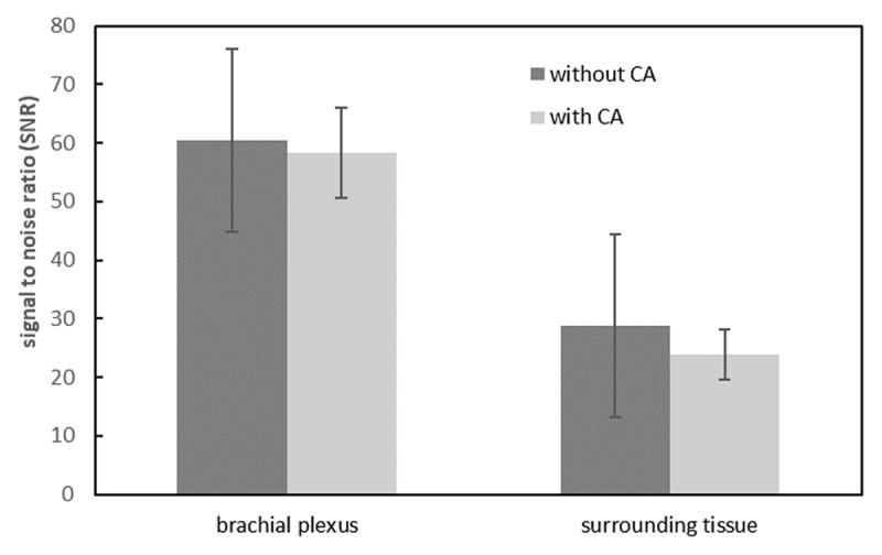
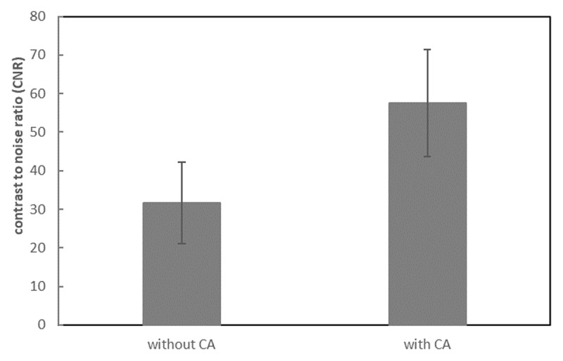
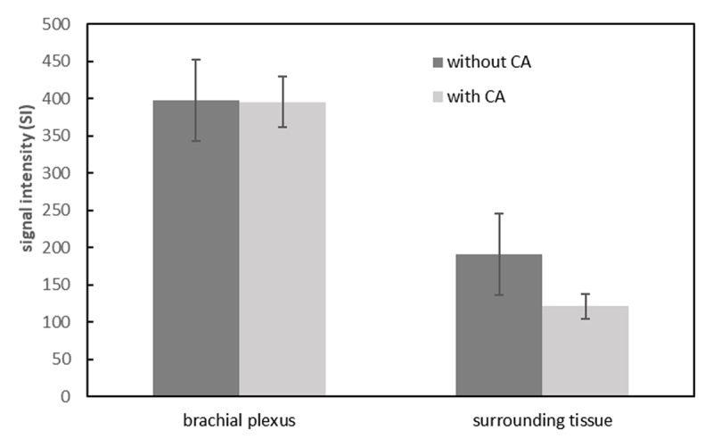
Results: The SNRs of the brachial plexus between 3D-STIR-VISTA without and with contrast agent were not significantly different, while SNRs of surrounding tissues were significantly decreased with contrast agent. The CNRs of 3D-STIR-VISTA images with contrast agent were significantly higher than that without contrast agent. The 3D-STIR-VISTA sequence with contrast agent exhibited a statistically higher CR than that without contrast agent. The average score for 3D-STIR-VISTA images with contrast agent was significantly higher than that without contrast agent.
Conclusion: The 3D-STIR-VISTA sequence with contrast agent is qualitatively and quantitatively superior to that without a contrast agent. The contrast-enhanced 3D-STIR-VISTA sequence can provide distinct visualization of the brachial plexus and enhance the contrast between the brachial plexus and surrounding tissues.
期刊介绍:
The purpose of the Journal of the Belgian Society of Radiology is the publication of articles dealing with diagnostic and interventional radiology, related imaging techniques, allied sciences, and continuing education.
 求助内容:
求助内容: 应助结果提醒方式:


