{"title":"脑蛛网膜下腔小梁的组织学和形态学。","authors":"Parisa Saboori, Ali Sadegh","doi":"10.1155/2015/279814","DOIUrl":null,"url":null,"abstract":"<p><p>The interface between the brain and the skull consists of three fibrous tissue layers, dura mater, arachnoid, and pia mater, known as the meninges, and strands of collagen tissues connecting the arachnoid to the pia mater, known as trabeculae. The space between the arachnoid and the pia mater is filled with cerebrospinal fluid which stabilizes the shape and position of the brain during head movements or impacts. The histology and architecture of the subarachnoid space trabeculae in the brain are not well established in the literature. The only recognized fact about the trabeculae is that they are made of collagen fibers surrounded by fibroblast cells and they have pillar- and veil-like structures. In this work the histology and the architecture of the brain trabeculae were studied, via a series of in vivo and in vitro experiments using cadaveric and animal tissue. In the cadaveric study fluorescence and bright field microscopy were employed while scanning and transmission electron microscopy were used for the animal studies. The results of this study reveal that the trabeculae are collagen based type I, and their architecture is in the form of tree-shaped rods, pillars, and plates and, in some regions, they have a complex network morphology. </p>","PeriodicalId":89526,"journal":{"name":"Anatomy research international","volume":"2015 ","pages":"279814"},"PeriodicalIF":0.0000,"publicationDate":"2015-01-01","publicationTypes":"Journal Article","fieldsOfStudy":null,"isOpenAccess":false,"openAccessPdf":"https://sci-hub-pdf.com/10.1155/2015/279814","citationCount":"53","resultStr":"{\"title\":\"Histology and Morphology of the Brain Subarachnoid Trabeculae.\",\"authors\":\"Parisa Saboori, Ali Sadegh\",\"doi\":\"10.1155/2015/279814\",\"DOIUrl\":null,\"url\":null,\"abstract\":\"<p><p>The interface between the brain and the skull consists of three fibrous tissue layers, dura mater, arachnoid, and pia mater, known as the meninges, and strands of collagen tissues connecting the arachnoid to the pia mater, known as trabeculae. The space between the arachnoid and the pia mater is filled with cerebrospinal fluid which stabilizes the shape and position of the brain during head movements or impacts. The histology and architecture of the subarachnoid space trabeculae in the brain are not well established in the literature. The only recognized fact about the trabeculae is that they are made of collagen fibers surrounded by fibroblast cells and they have pillar- and veil-like structures. In this work the histology and the architecture of the brain trabeculae were studied, via a series of in vivo and in vitro experiments using cadaveric and animal tissue. In the cadaveric study fluorescence and bright field microscopy were employed while scanning and transmission electron microscopy were used for the animal studies. The results of this study reveal that the trabeculae are collagen based type I, and their architecture is in the form of tree-shaped rods, pillars, and plates and, in some regions, they have a complex network morphology. </p>\",\"PeriodicalId\":89526,\"journal\":{\"name\":\"Anatomy research international\",\"volume\":\"2015 \",\"pages\":\"279814\"},\"PeriodicalIF\":0.0000,\"publicationDate\":\"2015-01-01\",\"publicationTypes\":\"Journal Article\",\"fieldsOfStudy\":null,\"isOpenAccess\":false,\"openAccessPdf\":\"https://sci-hub-pdf.com/10.1155/2015/279814\",\"citationCount\":\"53\",\"resultStr\":null,\"platform\":\"Semanticscholar\",\"paperid\":null,\"PeriodicalName\":\"Anatomy research international\",\"FirstCategoryId\":\"1085\",\"ListUrlMain\":\"https://doi.org/10.1155/2015/279814\",\"RegionNum\":0,\"RegionCategory\":null,\"ArticlePicture\":[],\"TitleCN\":null,\"AbstractTextCN\":null,\"PMCID\":null,\"EPubDate\":\"2015/5/24 0:00:00\",\"PubModel\":\"Epub\",\"JCR\":\"\",\"JCRName\":\"\",\"Score\":null,\"Total\":0}","platform":"Semanticscholar","paperid":null,"PeriodicalName":"Anatomy research international","FirstCategoryId":"1085","ListUrlMain":"https://doi.org/10.1155/2015/279814","RegionNum":0,"RegionCategory":null,"ArticlePicture":[],"TitleCN":null,"AbstractTextCN":null,"PMCID":null,"EPubDate":"2015/5/24 0:00:00","PubModel":"Epub","JCR":"","JCRName":"","Score":null,"Total":0}
Histology and Morphology of the Brain Subarachnoid Trabeculae.
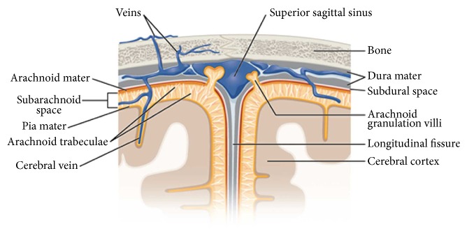
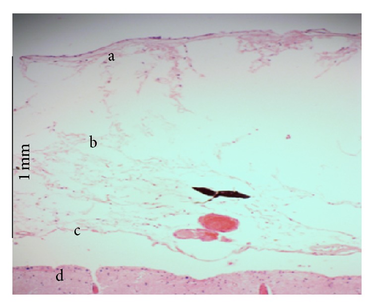
The interface between the brain and the skull consists of three fibrous tissue layers, dura mater, arachnoid, and pia mater, known as the meninges, and strands of collagen tissues connecting the arachnoid to the pia mater, known as trabeculae. The space between the arachnoid and the pia mater is filled with cerebrospinal fluid which stabilizes the shape and position of the brain during head movements or impacts. The histology and architecture of the subarachnoid space trabeculae in the brain are not well established in the literature. The only recognized fact about the trabeculae is that they are made of collagen fibers surrounded by fibroblast cells and they have pillar- and veil-like structures. In this work the histology and the architecture of the brain trabeculae were studied, via a series of in vivo and in vitro experiments using cadaveric and animal tissue. In the cadaveric study fluorescence and bright field microscopy were employed while scanning and transmission electron microscopy were used for the animal studies. The results of this study reveal that the trabeculae are collagen based type I, and their architecture is in the form of tree-shaped rods, pillars, and plates and, in some regions, they have a complex network morphology.

 求助内容:
求助内容: 应助结果提醒方式:


