Alexandra Papoudou-Bai, Alexandra Barbouti, Vassiliki Galani, Kalliopi Stefanaki, Panagiotis Kanavaros
{"title":"胸腺jun家族及转录信号转导和激活因子的免疫组织学分析。","authors":"Alexandra Papoudou-Bai, Alexandra Barbouti, Vassiliki Galani, Kalliopi Stefanaki, Panagiotis Kanavaros","doi":"10.1155/2015/541582","DOIUrl":null,"url":null,"abstract":"<p><p>The Jun family and the signal transducers and activators of transcription (STAT) are involved in proliferation and apoptosis. Moreover, c-Jun and STAT3 cooperate to regulate apoptosis. Therefore, we used double immunostaining to investigate the immunotopographical distribution of phospho-c-Jun (p-c-Jun), JunB, JunD, p-STAT3, p-STAT5, and p-STAT6 in human thymus. JunD was frequently expressed by thymocytes with higher expression in medullary compared to cortical thymocytes. p-c-Jun was frequently expressed by cortical and medullary thymic epithelial cells (TEC) and Hassall bodies (HB). p-STAT3 was frequently expressed by TEC with higher expression in cortical compared to medullary TEC and HB. p-c-Jun, JunB, p-STAT3, p-STAT5, and p-STAT6 were rarely expressed by thymocytes. JunB and JunD were expressed by rare cortical TEC with higher expression in medullary TEC. p-STAT5 and p-STAT6 were expressed by rare cortical and medullary TEC. Double immunostaining revealed p-c-Jun and JunD expression in rare CD11c positive dendritic cells. Our findings suggest a notable implication of JunD in the physiology of thymocytes and p-c-Jun and p-STAT3 in the physiology of TEC. The diversity of the immunotopographical distribution and the expression levels of p-c-Jun, JunB, JunD, p-STAT3, p-STAT5, and p-STAT6 indicates that they are differentially involved in the differentiation of TEC, thymocytes, and dendritic cells. </p>","PeriodicalId":89526,"journal":{"name":"Anatomy research international","volume":"2015 ","pages":"541582"},"PeriodicalIF":0.0000,"publicationDate":"2015-01-01","publicationTypes":"Journal Article","fieldsOfStudy":null,"isOpenAccess":false,"openAccessPdf":"https://www.ncbi.nlm.nih.gov/pmc/articles/PMC4381968/pdf/","citationCount":"2","resultStr":"{\"title\":\"Immunohistological analysis of the jun family and the signal transducers and activators of transcription in thymus.\",\"authors\":\"Alexandra Papoudou-Bai, Alexandra Barbouti, Vassiliki Galani, Kalliopi Stefanaki, Panagiotis Kanavaros\",\"doi\":\"10.1155/2015/541582\",\"DOIUrl\":null,\"url\":null,\"abstract\":\"<p><p>The Jun family and the signal transducers and activators of transcription (STAT) are involved in proliferation and apoptosis. Moreover, c-Jun and STAT3 cooperate to regulate apoptosis. Therefore, we used double immunostaining to investigate the immunotopographical distribution of phospho-c-Jun (p-c-Jun), JunB, JunD, p-STAT3, p-STAT5, and p-STAT6 in human thymus. JunD was frequently expressed by thymocytes with higher expression in medullary compared to cortical thymocytes. p-c-Jun was frequently expressed by cortical and medullary thymic epithelial cells (TEC) and Hassall bodies (HB). p-STAT3 was frequently expressed by TEC with higher expression in cortical compared to medullary TEC and HB. p-c-Jun, JunB, p-STAT3, p-STAT5, and p-STAT6 were rarely expressed by thymocytes. JunB and JunD were expressed by rare cortical TEC with higher expression in medullary TEC. p-STAT5 and p-STAT6 were expressed by rare cortical and medullary TEC. Double immunostaining revealed p-c-Jun and JunD expression in rare CD11c positive dendritic cells. Our findings suggest a notable implication of JunD in the physiology of thymocytes and p-c-Jun and p-STAT3 in the physiology of TEC. The diversity of the immunotopographical distribution and the expression levels of p-c-Jun, JunB, JunD, p-STAT3, p-STAT5, and p-STAT6 indicates that they are differentially involved in the differentiation of TEC, thymocytes, and dendritic cells. </p>\",\"PeriodicalId\":89526,\"journal\":{\"name\":\"Anatomy research international\",\"volume\":\"2015 \",\"pages\":\"541582\"},\"PeriodicalIF\":0.0000,\"publicationDate\":\"2015-01-01\",\"publicationTypes\":\"Journal Article\",\"fieldsOfStudy\":null,\"isOpenAccess\":false,\"openAccessPdf\":\"https://www.ncbi.nlm.nih.gov/pmc/articles/PMC4381968/pdf/\",\"citationCount\":\"2\",\"resultStr\":null,\"platform\":\"Semanticscholar\",\"paperid\":null,\"PeriodicalName\":\"Anatomy research international\",\"FirstCategoryId\":\"1085\",\"ListUrlMain\":\"https://doi.org/10.1155/2015/541582\",\"RegionNum\":0,\"RegionCategory\":null,\"ArticlePicture\":[],\"TitleCN\":null,\"AbstractTextCN\":null,\"PMCID\":null,\"EPubDate\":\"2015/3/18 0:00:00\",\"PubModel\":\"Epub\",\"JCR\":\"\",\"JCRName\":\"\",\"Score\":null,\"Total\":0}","platform":"Semanticscholar","paperid":null,"PeriodicalName":"Anatomy research international","FirstCategoryId":"1085","ListUrlMain":"https://doi.org/10.1155/2015/541582","RegionNum":0,"RegionCategory":null,"ArticlePicture":[],"TitleCN":null,"AbstractTextCN":null,"PMCID":null,"EPubDate":"2015/3/18 0:00:00","PubModel":"Epub","JCR":"","JCRName":"","Score":null,"Total":0}
Immunohistological analysis of the jun family and the signal transducers and activators of transcription in thymus.
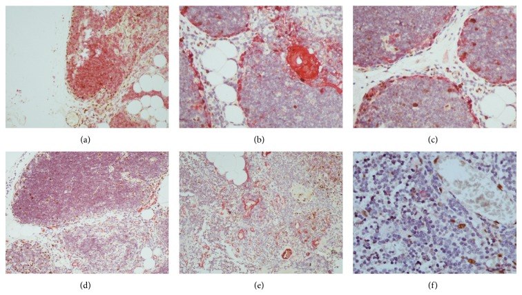
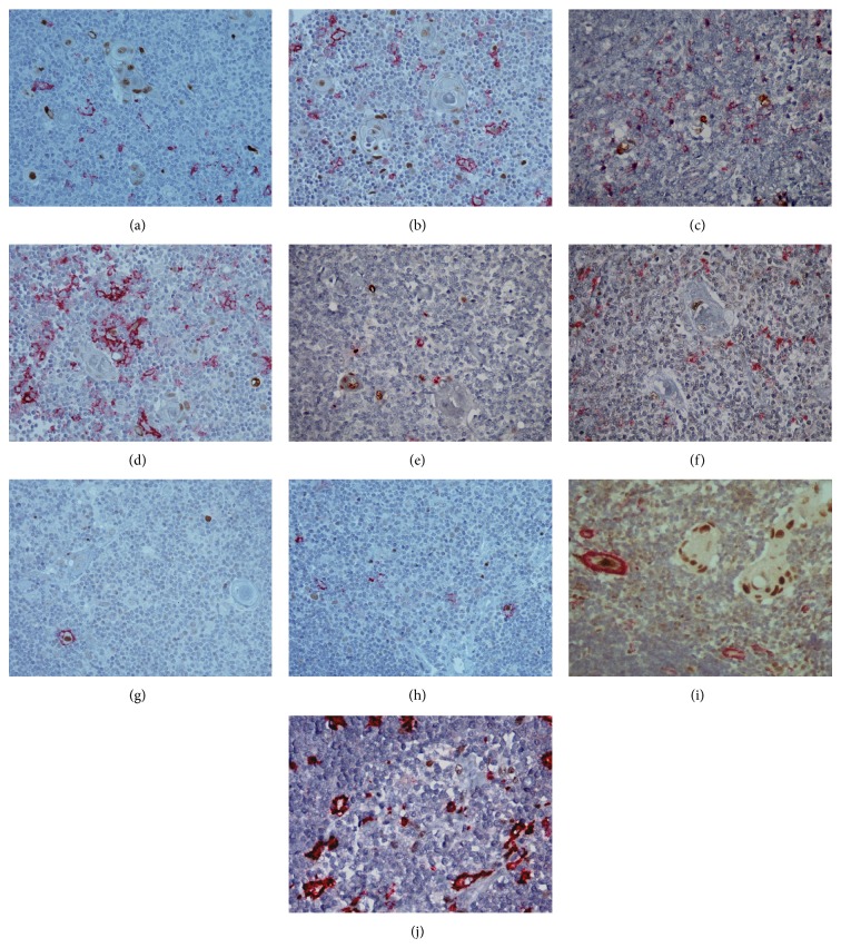
The Jun family and the signal transducers and activators of transcription (STAT) are involved in proliferation and apoptosis. Moreover, c-Jun and STAT3 cooperate to regulate apoptosis. Therefore, we used double immunostaining to investigate the immunotopographical distribution of phospho-c-Jun (p-c-Jun), JunB, JunD, p-STAT3, p-STAT5, and p-STAT6 in human thymus. JunD was frequently expressed by thymocytes with higher expression in medullary compared to cortical thymocytes. p-c-Jun was frequently expressed by cortical and medullary thymic epithelial cells (TEC) and Hassall bodies (HB). p-STAT3 was frequently expressed by TEC with higher expression in cortical compared to medullary TEC and HB. p-c-Jun, JunB, p-STAT3, p-STAT5, and p-STAT6 were rarely expressed by thymocytes. JunB and JunD were expressed by rare cortical TEC with higher expression in medullary TEC. p-STAT5 and p-STAT6 were expressed by rare cortical and medullary TEC. Double immunostaining revealed p-c-Jun and JunD expression in rare CD11c positive dendritic cells. Our findings suggest a notable implication of JunD in the physiology of thymocytes and p-c-Jun and p-STAT3 in the physiology of TEC. The diversity of the immunotopographical distribution and the expression levels of p-c-Jun, JunB, JunD, p-STAT3, p-STAT5, and p-STAT6 indicates that they are differentially involved in the differentiation of TEC, thymocytes, and dendritic cells.

 求助内容:
求助内容: 应助结果提醒方式:


