颞骨脑膜瘤表现为浆液性中耳炎1例。
Acta Radiologica Short Reports
Pub Date : 2014-11-26
eCollection Date: 2014-11-01
DOI:10.1177/2047981614555048
引用次数: 9
摘要
我们报告一例颞骨脑膜瘤延伸到中耳腔,临床表现为浆液性中耳炎的影像特征。颞骨脑膜瘤延伸至乳突或中耳腔,是非常罕见的。如果原因不明或治疗抵抗浆液性中耳炎和鼻咽肿瘤被排除,应进行颞骨计算机断层扫描(CT)。如果CT表现提示颞骨脑膜瘤,则磁共振成像(MRI)加钆检查将确认诊断并显示病变的确切范围。本文章由计算机程序翻译,如有差异,请以英文原文为准。
A case of a temporal bone meningioma presenting as a serous otitis media.
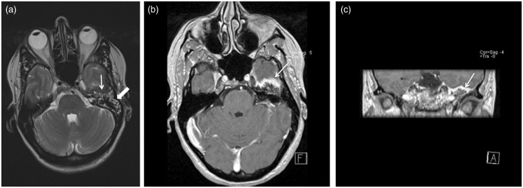
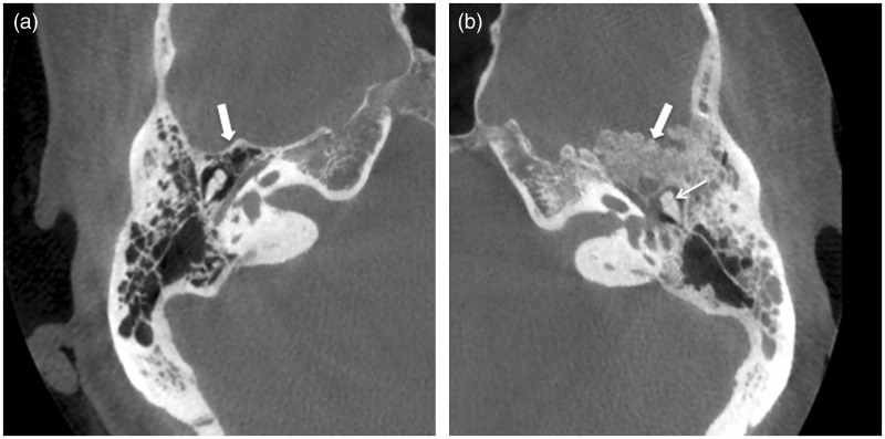
We report the imaging features of a case of a temporal bone meningioma extending into the middle ear cavity and clinically presenting as a serous otitis media. Temporal bone meningioma extending in the mastoid or the middle ear cavity, however, is very rare. In case of unexplained or therapy-resistant serous otitis media and a nasopharyngeal tumor being ruled out, a temporal bone computed tomography (CT) should be performed. If CT findings are suggestive of a temporal bone meningioma, a magnetic resonance imaging (MRI) examination with gadolinium will confirm diagnosis and show the exact extension of the lesion.
求助全文
通过发布文献求助,成功后即可免费获取论文全文。
去求助
来源期刊

自引率
0.00%
发文量
0
审稿时长
12 weeks
期刊介绍:
Under the editorial leadership of Professor Arnulf Skjennald MD and a distinguished editorial board, Acta Radiologica Open, formerly Acta Radiologica Short Reports, aims for the prompt publication of original case reports, short reports, review articles, pictorial reviews, research articles on diagnostic and interventional radiology, clinical radiology, experimental investigations in animals, and all other research related to imaging procedures. Acta Radiologica Open provides a complete update on all radiological specialties and technical utilities, as well as physiology and physics related to imaging, including ultrasonography, computed tomography, radionuclide and magnetic resonance imaging. Acta Radiologica Open publishes articles on diagnostic and interventional procedures in radiology based on all medical imaging techniques, as well as works in physiology and physics when related to radiology. The journal is an online-only, peer reviewed, open access journal.
 求助内容:
求助内容: 应助结果提醒方式:


